
nid: 58138
Additional formats:
None available
Description:
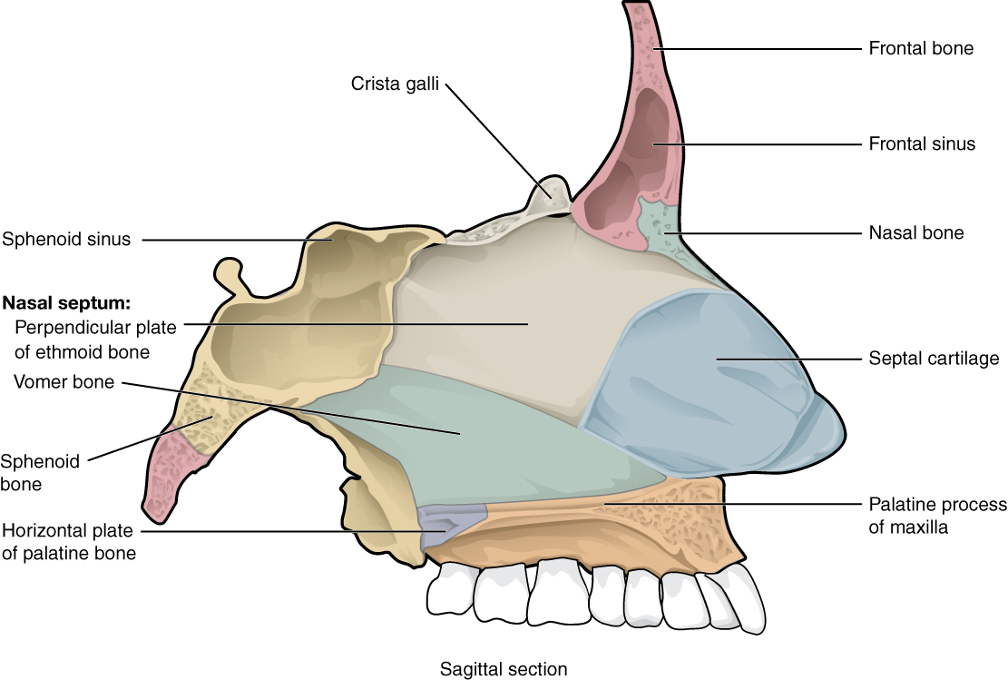
Nasal Septum. The nasal septum is formed by the perpendicular plate of the ethmoid bone and the vomer bone. The septal cartilage fills the gap between these bones and extends into the nose. English labels. From OpenStax book 'Anatomy and Physiology', fig. 7.17.
Anatomical structures in item:
Uploaded by: Jorn IJkhout
Netherlands, Leiden – Leiden University Medical Center, Leiden University
Bone
Septum nasi
Cranium
Os nasale
Sinus frontalis
Crista galli
Os sphenoidale
Processus palatinus maxillae
Lamina horizontalis ossis palatini
Vomer
Lamina perpendicularis ossis ethmoidalis
Creator(s)/credit: OpenStax
Requirements for usage
You are free to use this item if you follow the requirements of the license:  View license
View license If you use this item you should credit it as follows:
- For usage in print - copy and paste the line below:
- For digital usage (e.g. in PowerPoint, Impress, Word, Writer) - copy and paste the line below (optionally add the license icon):
"OpenStax AnatPhys fig.7.17 - Bones of Nasal Cavity - English labels" at AnatomyTOOL.org by OpenStax, license: Creative Commons Attribution. Source: book 'Anatomy and Physiology', https://openstax.org/details/books/anatomy-and-physiology.
"OpenStax AnatPhys fig.7.17 - Bones of Nasal Cavity - English labels" by OpenStax, license: CC BY. Source: book 'Anatomy and Physiology', https://openstax.org/details/books/anatomy-and-physiology.


Comments