
nid: 58487
Additional formats:
None available
Description:
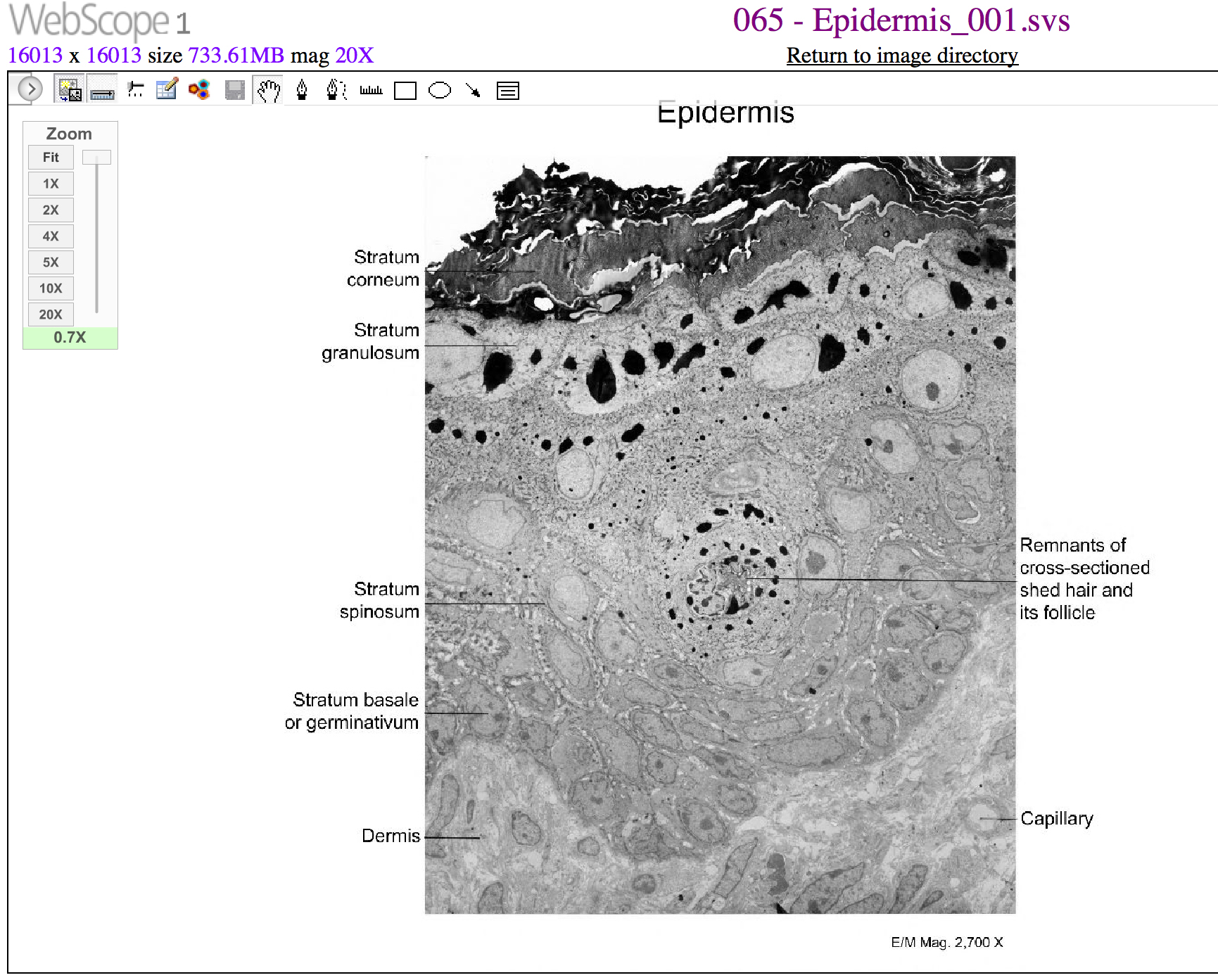
Cells of the Epidermis. The cells in the different layers of the epidermis originate from basal cells located in the stratum basale, yet the cells of each layer are distinctively different. EM × 2700. (Micrograph provided by the Regents of University of Michigan Medical School © 2012). English labels. From OpenStax book 'Anatomy and Physiology', fig. 5.6.
Anatomical structures in item:
Uploaded by: Jorn IJkhout
Netherlands, Leiden – Leiden University Medical Center, Leiden University
Epidermis
Dermis
Creator(s)/credit: OpenStax; Regents of U-M Medical School, UMich MedSchool
Requirements for usage
You are free to use this item if you follow the requirements of the license:  View license
View license If you use this item you should credit it as follows:
- For usage in print - copy and paste the line below:
- For digital usage (e.g. in PowerPoint, Impress, Word, Writer) - copy and paste the line below (optionally add the license icon):
"OpenStax AnatPhys fig.5.6 - Cells of the Epidermis - English labels" at AnatomyTOOL.org by OpenStax and Regents of U-M Medical School, UMich MedSchool, license: Creative Commons Attribution. Source: book 'Anatomy and Physiology', https://openstax.org/details/books/anatomy-and-physiology.
"OpenStax AnatPhys fig.5.6 - Cells of the Epidermis - English labels" by OpenStax and Regents of U-M Medical School, UMich MedSchool, license: CC BY. Source: book 'Anatomy and Physiology', https://openstax.org/details/books/anatomy-and-physiology.


Comments