
nid: 59290
Additional formats:
None available
Description:
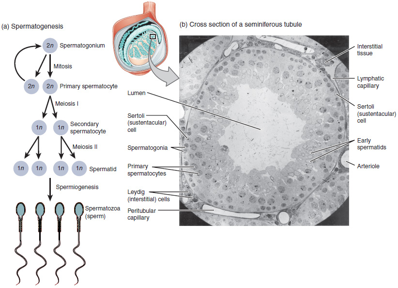
Spermatogenesis. (a) Mitosis of a spermatogonial stem cell involves a single cell division that results in two identical, diploid daughter cells (spermatogonia to primary spermatocyte). Meiosis has two rounds of cell division: primary spermatocyte to secondary spermatocyte, and then secondary spermatocyte to spermatid. This produces four haploid daughter cells (spermatids). (b) In this electron micrograph of a cross-section of a seminiferous tubule from a rat, the lumen is the light-shaded area in the center of the image. The location of the primary spermatocytes is near the basement membrane, and the early spermatids are approaching the lumen (tissue source: rat). EM × 900. (Micrograph provided by the Regents of University of Michigan Medical School © 2012). English labels. From OpenStax book 'Anatomy and Physiology', fig. 27.5.
Anatomical structures in item:
Uploaded by: Jorn IJkhout
Netherlands, Leiden – Leiden University Medical Center, Leiden University
Colliculus inferior
Tubuli seminiferi contorti
Creator(s)/credit: OpenStax; Regents of U-M Medical School, UMich MedSchool
Requirements for usage
You are free to use this item if you follow the requirements of the license:  View license
View license If you use this item you should credit it as follows:
- For usage in print - copy and paste the line below:
- For digital usage (e.g. in PowerPoint, Impress, Word, Writer) - copy and paste the line below (optionally add the license icon):
"OpenStax AnatPhys fig.27.5 - Spermatogenesis - English labels" at AnatomyTOOL.org by OpenStax and Regents of U-M Medical School, UMich MedSchool, license: Creative Commons Attribution. Source: book 'Anatomy and Physiology', https://openstax.org/details/books/anatomy-and-physiology.
"OpenStax AnatPhys fig.27.5 - Spermatogenesis - English labels" by OpenStax and Regents of U-M Medical School, UMich MedSchool, license: CC BY. Source: book 'Anatomy and Physiology', https://openstax.org/details/books/anatomy-and-physiology.


Comments