
nid: 59279
Additional formats:
None available
Description:
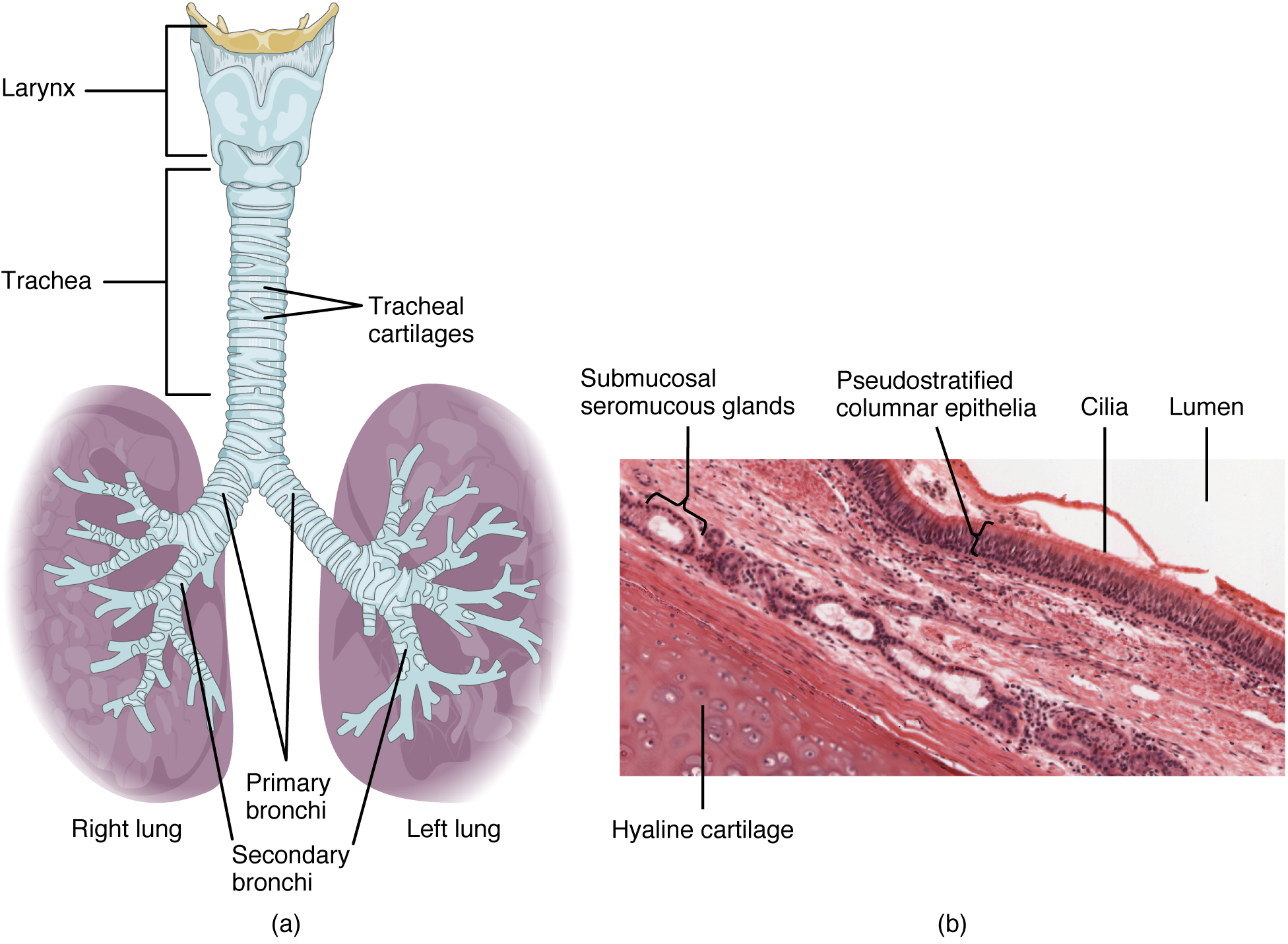
Trachea. (a) The tracheal tube is formed by stacked, C-shaped pieces of hyaline cartilage. (b) The layer visible in this cross-section of tracheal wall tissue between the hyaline cartilage and the lumen of the trachea is the mucosa, which is composed of pseudostratified ciliated columnar epithelium that contains goblet cells. LM × 1220. (Micrograph provided by the Regents of University of Michigan Medical School © 2012). English labels. From OpenStax book 'Anatomy and Physiology', fig. 22.9.
Anatomical structures in item:
Uploaded by: Jorn IJkhout
Netherlands, Leiden – Leiden University Medical Center, Leiden University
Systema respiratorium
Trachea
Larynx
Cartilagines tracheales
Cilia
Cilia
Bronchi
Pulmones
Creator(s)/credit: OpenStax; Regents of U-M Medical School, UMich MedSchool
Requirements for usage
You are free to use this item if you follow the requirements of the license:  View license
View license If you use this item you should credit it as follows:
- For usage in print - copy and paste the line below:
- For digital usage (e.g. in PowerPoint, Impress, Word, Writer) - copy and paste the line below (optionally add the license icon):
"OpenStax AnatPhys fig.22.9 - The Trachea - English labels" at AnatomyTOOL.org by OpenStax and Regents of U-M Medical School, UMich MedSchool, license: Creative Commons Attribution. Source: book 'Anatomy and Physiology', https://openstax.org/details/books/anatomy-and-physiology.
"OpenStax AnatPhys fig.22.9 - The Trachea - English labels" by OpenStax and Regents of U-M Medical School, UMich MedSchool, license: CC BY. Source: book 'Anatomy and Physiology', https://openstax.org/details/books/anatomy-and-physiology.


Comments