
nid: 59277
Additional formats:
None available
Description:
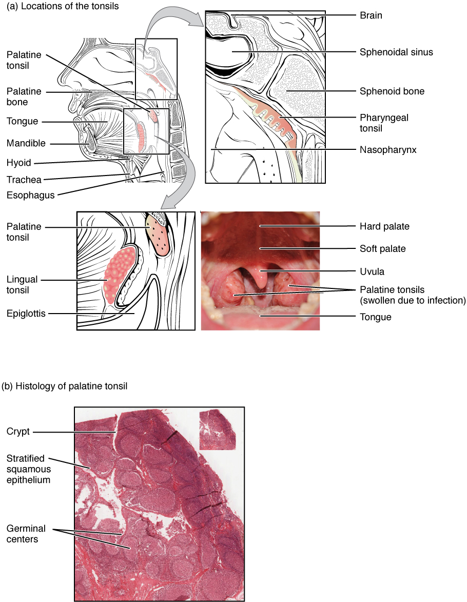
Locations and Histology of the Tonsils. (a) The pharyngeal tonsilis located on the roof of the posterior superior wall of the nasopharynx. The palatine tonsils lay on each side of the pharynx. (b) A micrograph shows the palatine tonsil tissue. LM × 40. (Micrograph provided by the Regents of the University of Michigan Medical School © 2012). English labels. From OpenStax book 'Anatomy and Physiology', fig. 21.10.
Anatomical structures in item:
Uploaded by: Jorn IJkhout
Netherlands, Leiden – Leiden University Medical Center, Leiden University
Tonsilla palatina
Tonsilla lingualis
Epiglottis
Palatum durum
Palatum molle
Uvula palatina
Tonsilla pharyngealis
Sinus sphenoidalis
Os sphenoidale
Os palatinum
Lingua
Mandibula
Os hyoideum
Trachea
Oesophagus
Creator(s)/credit: OpenStax; Regents of U-M Medical School, UMich MedSchool
Requirements for usage
You are free to use this item if you follow the requirements of the license:  View license
View license If you use this item you should credit it as follows:
- For usage in print - copy and paste the line below:
- For digital usage (e.g. in PowerPoint, Impress, Word, Writer) - copy and paste the line below (optionally add the license icon):
"OpenStax AnatPhys fig.21.10 - Location and History of Tonsils - English labels" at AnatomyTOOL.org by OpenStax and Regents of U-M Medical School, UMich MedSchool, license: Creative Commons Attribution. Source: book 'Anatomy and Physiology', https://openstax.org/details/books/anatomy-and-physiology.
"OpenStax AnatPhys fig.21.10 - Location and History of Tonsils - English labels" by OpenStax and Regents of U-M Medical School, UMich MedSchool, license: CC BY. Source: book 'Anatomy and Physiology', https://openstax.org/details/books/anatomy-and-physiology.


Comments