
nid: 58937
Additional formats:
None available
Description:
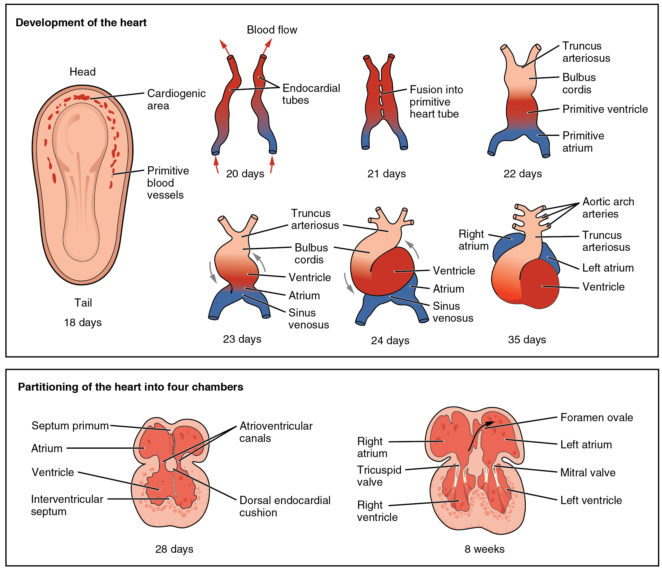
Development of the Human Heart. This diagram outlines the embryological development of the human heart during the first eight weeks and the subsequent formation of the four heart chambers. English labels. From OpenStax book 'Anatomy and Physiology', fig. 19.36.
Anatomical structures in item:
Uploaded by: Jorn IJkhout
Netherlands, Leiden – Leiden University Medical Center, Leiden University
Cor
Sinus venosus
Atrium sinistrum
Atrium dextrum
Arcus aortae
Septum interventriculare
Ventriculus dexter
Ventriculus sinister
Valva tricuspidalis
Valva mitralis
Foramen ovale
Creator(s)/credit: OpenStax
Requirements for usage
You are free to use this item if you follow the requirements of the license:  View license
View license If you use this item you should credit it as follows:
- For usage in print - copy and paste the line below:
- For digital usage (e.g. in PowerPoint, Impress, Word, Writer) - copy and paste the line below (optionally add the license icon):
"OpenStax AnatPhys fig.19.36 - Embryonic Development of Heart - English labels" at AnatomyTOOL.org by OpenStax, license: Creative Commons Attribution. Source: book 'Anatomy and Physiology', https://openstax.org/details/books/anatomy-and-physiology.
"OpenStax AnatPhys fig.19.36 - Embryonic Development of Heart - English labels" by OpenStax, license: CC BY. Source: book 'Anatomy and Physiology', https://openstax.org/details/books/anatomy-and-physiology.


Comments