
nid: 59269
Additional formats:
None available
Description:
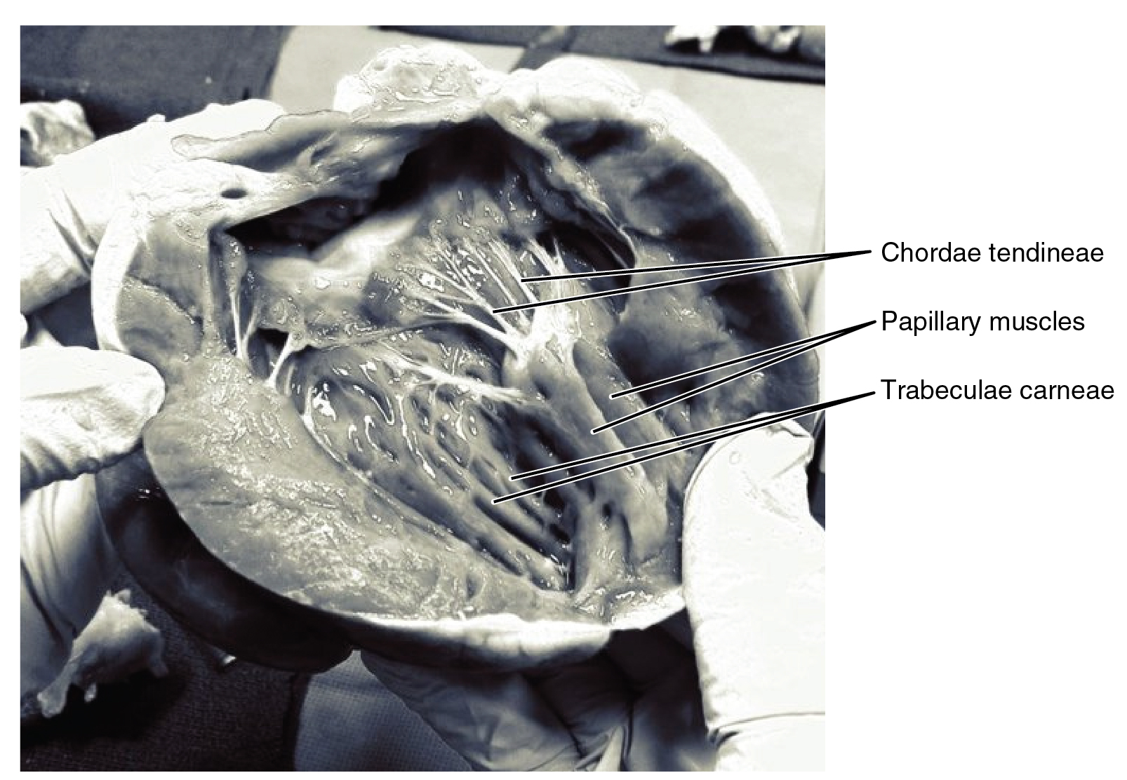
Chordae Tendineae and Papillary Muscles. In this frontal section, you can see papillary muscles attached to the tricuspid valve on the right as well as the mitral valve on the left via chordae tendineae. (credit: modification of work by “PV KS”/flickr.com). English labels. From OpenStax book 'Anatomy and Physiology', fig. 19.11.
Anatomical structures in item:
Uploaded by: Jorn IJkhout
Netherlands, Leiden – Leiden University Medical Center, Leiden University
Cor
Chordae tendineae cordis
Musculi papillares cordis
Trabeculae carneae cordis
Valva tricuspidalis
Creator(s)/credit: OpenStax; PV KS
Requirements for usage
You are free to use this item if you follow the requirements of the license:  View license
View license If you use this item you should credit it as follows:
- For usage in print - copy and paste the line below:
- For digital usage (e.g. in PowerPoint, Impress, Word, Writer) - copy and paste the line below (optionally add the license icon):
"OpenStax AnatPhys fig.19.11 - Chordae Tendinae Papillary Muscles - English labels" at AnatomyTOOL.org by OpenStax and PV KS, license: Creative Commons Attribution. Source: book 'Anatomy and Physiology', https://openstax.org/details/books/anatomy-and-physiology.
"OpenStax AnatPhys fig.19.11 - Chordae Tendinae Papillary Muscles - English labels" by OpenStax and PV KS, license: CC BY. Source: book 'Anatomy and Physiology', https://openstax.org/details/books/anatomy-and-physiology.


Comments