
nid: 58813
Additional formats:
None available
Description:
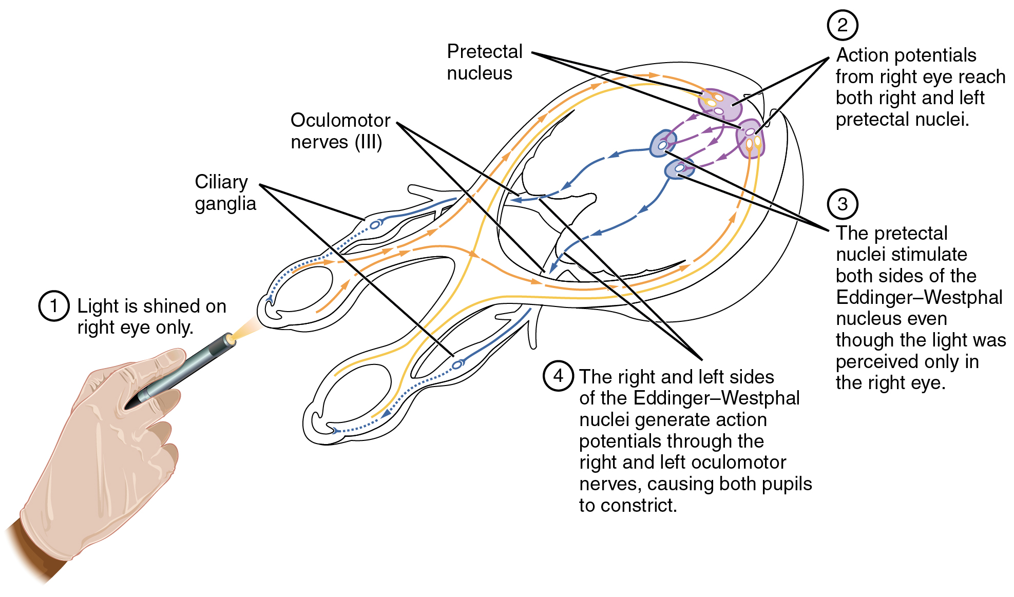
Pupillary Reflex Pathways. The pupil is under competing autonomic control in response to light levels hitting the retina. The sympathetic system will dilate the pupil when the retina is not receiving enough light, and the parasympathetic system will constrict the pupil when too much light hits the retina. English labels. From OpenStax book 'Anatomy and Physiology', fig. 15.10.
Anatomical structures in item:
Uploaded by: Jorn IJkhout
Netherlands, Leiden – Leiden University Medical Center, Leiden University
Encephalon
Pupilla
Ganglion ciliare
Nervus opticus
Nervus oculomotorius [III]
Area pretectalis
Creator(s)/credit: OpenStax
Requirements for usage
You are free to use this item if you follow the requirements of the license:  View license
View license If you use this item you should credit it as follows:
- For usage in print - copy and paste the line below:
- For digital usage (e.g. in PowerPoint, Impress, Word, Writer) - copy and paste the line below (optionally add the license icon):
"OpenStax AnatPhys fig.15.10 - Pupillary Reflex Pathways - English labels" at AnatomyTOOL.org by OpenStax, license: Creative Commons Attribution. Source: book 'Anatomy and Physiology', https://openstax.org/details/books/anatomy-and-physiology.
"OpenStax AnatPhys fig.15.10 - Pupillary Reflex Pathways - English labels" by OpenStax, license: CC BY. Source: book 'Anatomy and Physiology', https://openstax.org/details/books/anatomy-and-physiology.


Comments