
nid: 58743
Additional formats:
None available
Description:
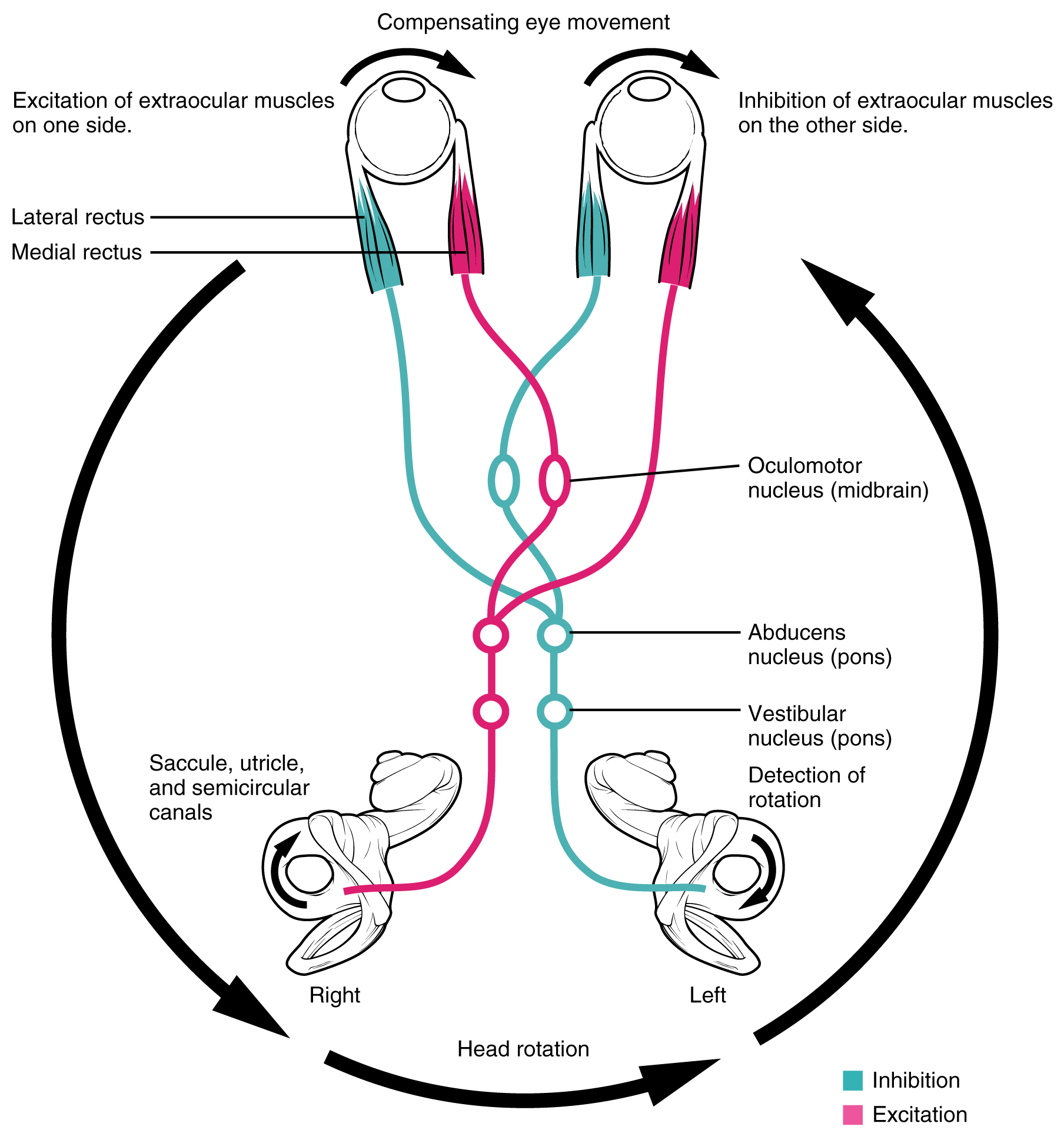
Vestibulo-ocular Reflex. Connections between the vestibular system and the cranial nerves controlling eye movement keep the eyes centered on a visual stimulus, even though the head is moving. During head movement, the eye muscles move the eyes in the opposite direction as the head movement, keeping the visual stimulus centered in the field of view. English labels. From OpenStax book 'Anatomy and Physiology', fig. 14.21.
Anatomical structures in item:
Uploaded by: Jorn IJkhout
Netherlands, Leiden – Leiden University Medical Center, Leiden University
Encephalon
Oculus
Musculus rectus lateralis
Musculus rectus medialis
Nucleus nervi oculomotorii
Nucleus nervi abducentis
Nuclei vestibulares in tegmento pontis
Sacculus
Utriculus
Canales semicirculares
Creator(s)/credit: OpenStax
Requirements for usage
You are free to use this item if you follow the requirements of the license:  View license
View license If you use this item you should credit it as follows:
- For usage in print - copy and paste the line below:
- For digital usage (e.g. in PowerPoint, Impress, Word, Writer) - copy and paste the line below (optionally add the license icon):
"OpenStax AnatPhys fig.14.21 - Vestibulo-Ocular Reflex - English labels" at AnatomyTOOL.org by OpenStax, license: Creative Commons Attribution. Source: book 'Anatomy and Physiology', https://openstax.org/details/books/anatomy-and-physiology.
"OpenStax AnatPhys fig.14.21 - Vestibulo-Ocular Reflex - English labels" by OpenStax, license: CC BY. Source: book 'Anatomy and Physiology', https://openstax.org/details/books/anatomy-and-physiology.


Comments