
nid: 59262
Additional formats:
None available
Description:
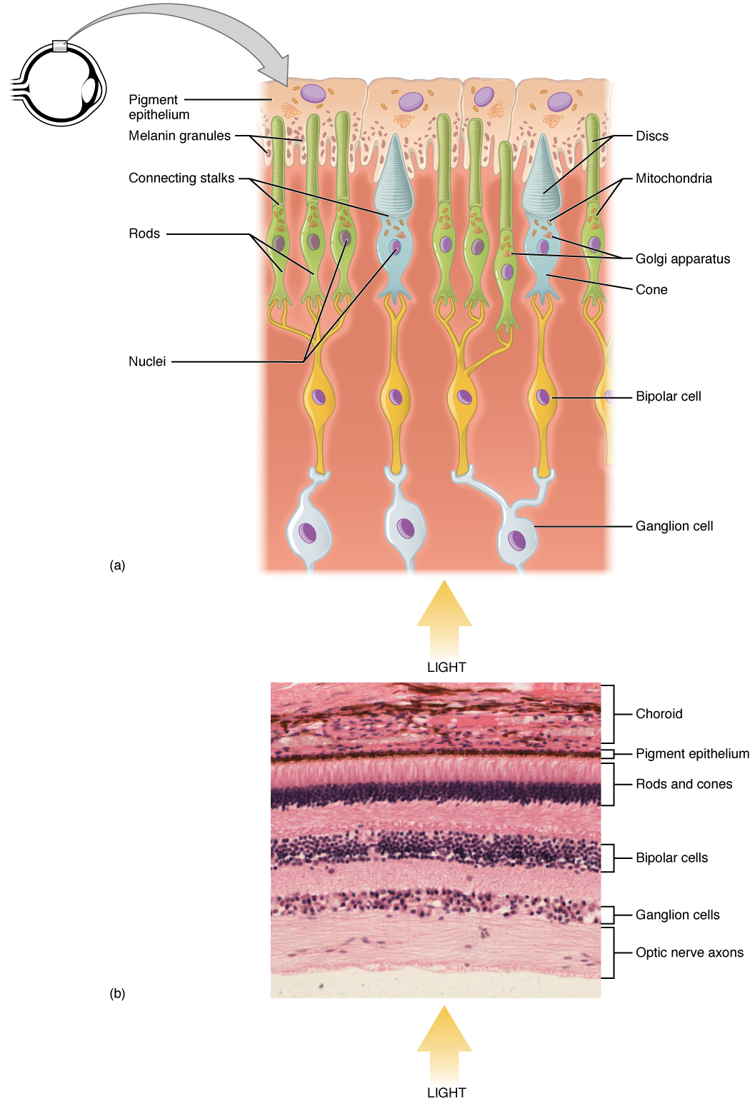
Photoreceptor. (a) All photoreceptors have inner segments containing the nucleus and other important organelles and outer segments with membrane arrays containing the photosensitive opsin molecules. Rod outer segments are long columnar shapes with stacks of membrane-bound discs that contain the rhodopsin pigment. Cone outer segments are short, tapered shapes with folds of membrane in place of the discs in the rods. (b) Tissue of the retina shows a dense layer of nuclei of the rods and cones. LM × 800. (Micrograph provided by the Regents of University of Michigan Medical School © 2012). English labels. From OpenStax book 'Anatomy and Physiology', fig. 14.16.
Anatomical structures in item:
Uploaded by: Jorn IJkhout
Netherlands, Leiden – Leiden University Medical Center, Leiden University
Stratum segmentorum externorum et internorum retinae
Oculus
Choroidea
Stratum pigmentosum retinae
Stratum ganglionicum retinae
Nervus opticus
Creator(s)/credit: OpenStax; Regents of U-M Medical School, UMich MedSchool
Requirements for usage
You are free to use this item if you follow the requirements of the license:  View license
View license If you use this item you should credit it as follows:
- For usage in print - copy and paste the line below:
- For digital usage (e.g. in PowerPoint, Impress, Word, Writer) - copy and paste the line below (optionally add the license icon):
"OpenStax AnatPhys fig.14.16 - Rods and Cones - English labels" at AnatomyTOOL.org by OpenStax and Regents of U-M Medical School, UMich MedSchool, license: Creative Commons Attribution. Source: book 'Anatomy and Physiology', https://openstax.org/details/books/anatomy-and-physiology.
"OpenStax AnatPhys fig.14.16 - Rods and Cones - English labels" by OpenStax and Regents of U-M Medical School, UMich MedSchool, license: CC BY. Source: book 'Anatomy and Physiology', https://openstax.org/details/books/anatomy-and-physiology.


Comments