
nid: 59256
Additional formats:
None available
Description:
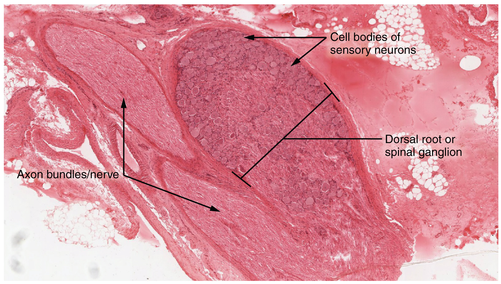
Dorsal Root Ganglion. The cell bodies of sensory neurons, which are unipolar neurons by shape, are seen in this photomicrograph. Also, the fibrous region is composed of the axons of these neurons that are passing through the ganglion to be part of the dorsal nerve root (tissue source: canine). LM × 40. (Micrograph provided by the Regents of University of Michigan Medical School © 2012). English labels. From OpenStax book 'Anatomy and Physiology', fig. 13.19.
Anatomical structures in item:
Uploaded by: Jorn IJkhout
Netherlands, Leiden – Leiden University Medical Center, Leiden University
Neuron
Ganglion sensorium nervi spinalis
Creator(s)/credit: OpenStax; Regents of U-M Medical School, UMich MedSchool
Requirements for usage
You are free to use this item if you follow the requirements of the license:  View license
View license If you use this item you should credit it as follows:
- For usage in print - copy and paste the line below:
- For digital usage (e.g. in PowerPoint, Impress, Word, Writer) - copy and paste the line below (optionally add the license icon):
"OpenStax AnatPhys fig.13.19 - Dorsal Root Ganglion - English labels" at AnatomyTOOL.org by OpenStax and Regents of U-M Medical School, UMich MedSchool, license: Creative Commons Attribution. Source: book 'Anatomy and Physiology', https://openstax.org/details/books/anatomy-and-physiology.
"OpenStax AnatPhys fig.13.19 - Dorsal Root Ganglion - English labels" by OpenStax and Regents of U-M Medical School, UMich MedSchool, license: CC BY. Source: book 'Anatomy and Physiology', https://openstax.org/details/books/anatomy-and-physiology.


Comments