
nid: 58673
Additional formats:
None available
Description:
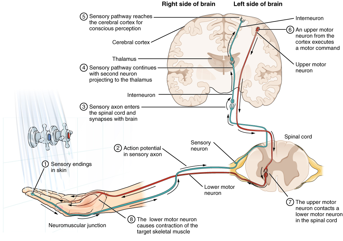
Testing the Water (1) The sensory neuron has endings in the skin that sense a stimulus such as water temperature. The strength of the signal that starts here is dependent on the strength of the stimulus. (2) The graded potential from the sensory endings, if strong enough, will initiate an action potential at the initial segment of the axon (which is immediately adjacent to the sensory endings in the skin). (3) The axon of the peripheral sensory neuron enters the spinal cord and contacts another neuron in the gray matter. The contact is a synapse where another graded potential is caused by the release of a chemical signal from the axon terminals. (4) An action potential is initiated at the initial segment of this neuron and travels up the sensory pathway to a region of the brain called the thalamus. Another synapse passes the information along to the next neuron. (5) The sensory pathway ends when the signal reaches the cerebral cortex. (6) After integration with neurons in other parts of the cerebral cortex, a motor command is sent from the precentral gyrus of the frontal cortex. (7) The upper motor neuron sends an action potential down to the spinal cord. The target of the upper motor neuron is the dendrites of the lower motor neuron in the gray matter of the spinal cord. (8) The axon of the lower motor neuron emerges from the spinal cord in a nerve and connects to a muscle through a neuromuscular junction to cause contraction of the target muscle. English labels. From OpenStax book 'Anatomy and Physiology', fig. 12.14.
Anatomical structures in item:
Uploaded by: Jorn IJkhout
Netherlands, Leiden – Leiden University Medical Center, Leiden University
Neuron
Nervus sensorius
Nervus motorius
Medulla spinalis
Thalamus
Cortex cerebelli
Creator(s)/credit: OpenStax
Requirements for usage
You are free to use this item if you follow the requirements of the license:  View license
View license If you use this item you should credit it as follows:
- For usage in print - copy and paste the line below:
- For digital usage (e.g. in PowerPoint, Impress, Word, Writer) - copy and paste the line below (optionally add the license icon):
"OpenStax AnatPhys fig.12.14 - Sensory Neuron Test Water - English labels " at AnatomyTOOL.org by OpenStax, license: Creative Commons Attribution. Source: book 'Anatomy and Physiology', https://openstax.org/details/books/anatomy-and-physiology.
"OpenStax AnatPhys fig.12.14 - Sensory Neuron Test Water - English labels " by OpenStax, license: CC BY. Source: book 'Anatomy and Physiology', https://openstax.org/details/books/anatomy-and-physiology.


Comments