
nid: 62480
Additional formats:
None available
Description:
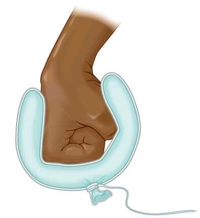
The balloon metaphor is often used to explain the pericardium, pleura and peritoneum. These are so-called serous membranes. They line the inner side of the body wall and reflect back to cover an organ, respectively: the heart, te lungs and the intestines — much the same way that an underinflated balloon would form two layers surrounding a fist.
The layer covering the organ is called the visceral layer (viscus = internal organ), the layer lining the inside of the outer wall is called the parietal layer (paries= wall).
Between the layers is a cavity. The cavity is collapsed. It only contains a little amount of fluid, serous fluid.
This 'construction' allows both layers to slide over each other, hence the surrounded organ to move or expand (a bit), whilst it is also secured in place. This 'construction' is seen around organs that need to move or expand.
Extracted from OpenStax book 'Anatomy and Physiology', fig. 1.17.
Anatomical structures in item:
Uploaded by: opgobee
Netherlands, Leiden – Leiden University Medical Center, Leiden University
Pericardium
Epicardium
Lamina parietalis pericardii
Cavitas pericardiaca
Peritoneum
Peritoneum parietale
Peritoneum viscerale
Pleura
Pleura parietalis
Pleura visceralis
Cavitas peritonealis
Cavitas pleuralis
Lamina visceralis
Creator(s)/credit: OpenStax
Requirements for usage
You are free to use this item if you follow the requirements of the license:  View license
View license If you use this item you should credit it as follows:
- For usage in print - copy and paste the line below:
- For digital usage (e.g. in PowerPoint, Impress, Word, Writer) - copy and paste the line below (optionally add the license icon):
"OpenStax AnatPhys fig.1.17 - Serous Membrane Balloon Metaphor - no labels" at AnatomyTOOL.org by OpenStax, license: Creative Commons Attribution. Source: book 'Anatomy and Physiology', https://openstax.org/details/books/anatomy-and-physiology.
"OpenStax AnatPhys fig.1.17 - Serous Membrane Balloon Metaphor - no labels" by OpenStax, license: CC BY. Source: book 'Anatomy and Physiology', https://openstax.org/details/books/anatomy-and-physiology.


Comments