
nid: 58578
Additional formats:
None available
Description:
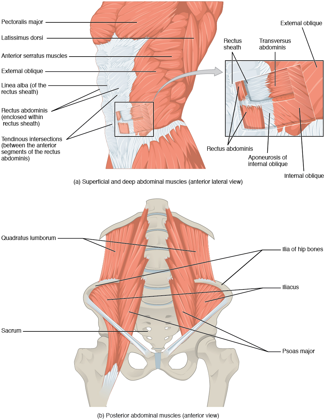
Muscles of the Abdomen. (a) The anterior abdominal muscles include the medially located rectus abdominis, which is covered by a sheet of connective tissue called the rectus sheath. On the flanks of the body, medial to the rectus abdominis, the abdominal wall is composed of three layers. The external oblique muscles form the superficial layer, while the internal oblique muscles form the middle layer, and the transverses abdominus forms the deepest layer. (b) The muscles of the lower back move the lumbar spine but also assist in femur movements. English labels. From OpenStax book 'Anatomy and Physiology', fig. 11.16.
Anatomical structures in item:
Uploaded by: Jorn IJkhout
Netherlands, Leiden – Leiden University Medical Center, Leiden University
Musculus pectoralis major
Musculus latissimus dorsi
Musculus serratus anterior
Musculus obliquus externus abdominis
Linea alba
Musculus rectus abdominis
Musculus transversus abdominis
Musculus quadratus lumborum
Os sacrum [vertebrae sacrales I - V]
Musculus psoas major
Musculus iliacus
Crista iliaca
Creator(s)/credit: OpenStax
Requirements for usage
You are free to use this item if you follow the requirements of the license:  View license
View license If you use this item you should credit it as follows:
- For usage in print - copy and paste the line below:
- For digital usage (e.g. in PowerPoint, Impress, Word, Writer) - copy and paste the line below (optionally add the license icon):
"OpenStax AnatPhys fig.11.16 - Muscles of the Abdomen - English labels" at AnatomyTOOL.org by OpenStax, license: Creative Commons Attribution. Source: book 'Anatomy and Physiology', https://openstax.org/details/books/anatomy-and-physiology.
"OpenStax AnatPhys fig.11.16 - Muscles of the Abdomen - English labels" by OpenStax, license: CC BY. Source: book 'Anatomy and Physiology', https://openstax.org/details/books/anatomy-and-physiology.


Comments