
nid: 60843
Additional formats:
None available
Description:
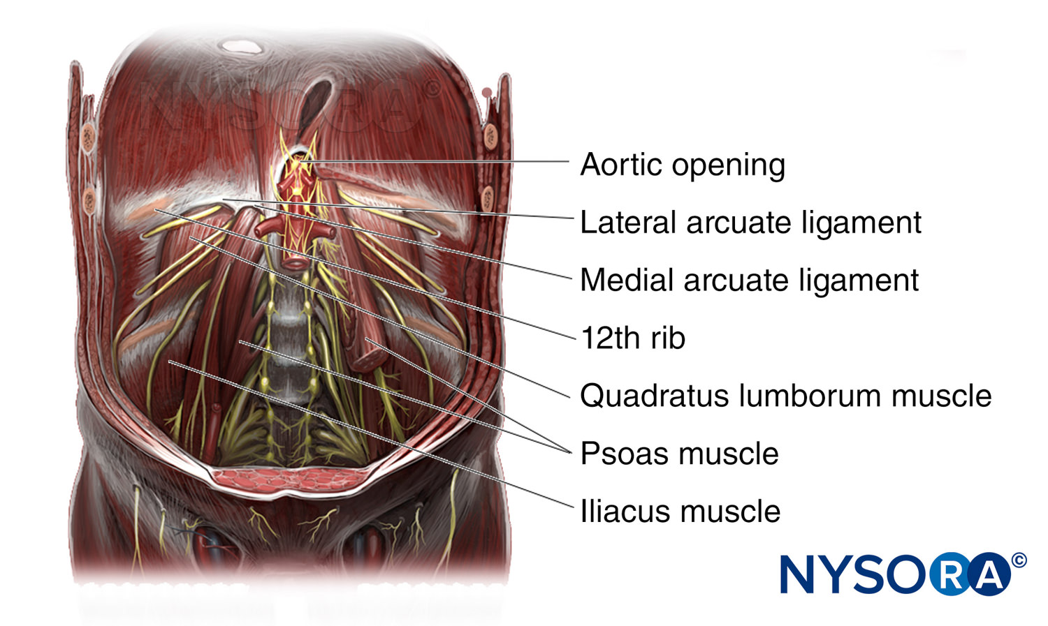
Posterior wall of abdomen. Certain structures of the posterior wall of the abdomen are showed. English labels.
Image created for NYSORA by VisionExpo.Design - www.nysora.com
Image created for NYSORA by VisionExpo.Design - www.nysora.com
Anatomical structures in item:
Uploaded by: rva
Netherlands, Leiden – Leiden University Medical Center, Leiden University
Abdomen
Hiatus aorticus
Ligamentum arcuatum laterale
Ligamentum arcuatum mediale
Musculus quadratus lumborum
Musculus psoas major
Musculus iliacus
Crus dextrum (Diaphragma)
Musculus psoas minor
Foramen venae cavae
Hiatus oesophageus
Diaphragma
Vertebrae lumbales (LI-LV)
Aorta abdominalis
Creator(s)/credit: New York School of Regional Anesthesia; VisionExpo.Design, illustration creation
Requirements for usage
You are free to use this item if you follow the requirements of the license:  View license
View license If you use this item you should credit it as follows:
- For usage in print - copy and paste the line below:
- For digital usage (e.g. in PowerPoint, Impress, Word, Writer) - copy and paste the line below (optionally add the license icon):
"NYSORA - Drawing Posterior wall of abdomen - English labels" at AnatomyTOOL.org by New York School of Regional Anesthesia and VisionExpo.Design, license: Creative Commons Attribution-NonCommercial-NoDerivs


Comments