nid: 60846
Additional formats:
None available
Description:
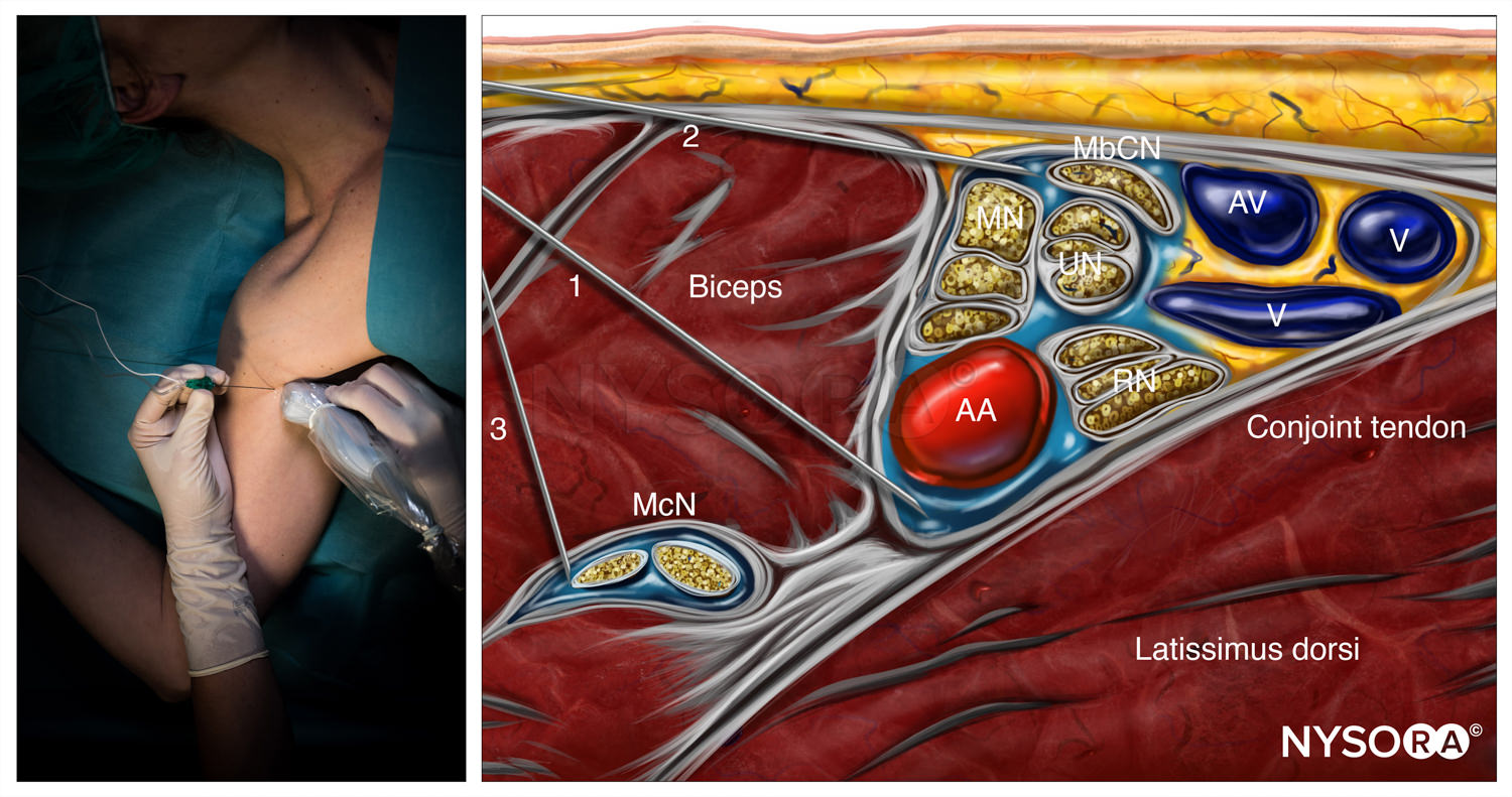
Axillary Brachial Plexus Block. This image shows the placement of needles in Axillary Brachial Plexus Block regional anesthesia, this technique is used is to deposit anesthetics locally around the axillary artery. Several nerves, veins, muscles and the axillary artery can be seen in the right image. In the left image the placement of the ultrasound device is shown. McN = musculocutaneous nerve, MN = median nerve, UN = ulnar nerve, RN = radial nerve, MbCN = medial brachial cutaneous nerve, AA = axillary artery, AV = axillary vein, and V = vein. English labels.
Image created for NYSORA by VisionExpo.Design - www.nysora.com
Image created for NYSORA by VisionExpo.Design - www.nysora.com
Anatomical structures in item:
Uploaded by: rva
Netherlands, Leiden – Leiden University Medical Center, Leiden University
Musculus biceps brachii
Nervus musculocutaneus
Arteria axillaris
Vena axillaris
Nervus medianus
Nervus ulnaris
Nervus radialis
Nervus cutaneus brachii medialis
Tendo conjunctivus
Musculus latissimus dorsi
Creator(s)/credit: New York School of Regional Anesthesia; VisionExpo.Design, illustration creation
Requirements for usage
You are free to use this item if you follow the requirements of the license:  View license
 View license
 If you use this item you should credit it as follows:
- For usage in print - copy and paste the line below:
- For digital usage (e.g. in PowerPoint, Impress, Word, Writer) - copy and paste the line below (optionally add the license icon):
"NYSORA - Drawing Axillary Brachial Plexus Block - English labels" at AnatomyTOOL.org by New York School of Regional Anesthesia and VisionExpo.Design, license: Creative Commons Attribution-NonCommercial-NoDerivs


Comments