nid: 60845
Additional formats:
None available
Description:
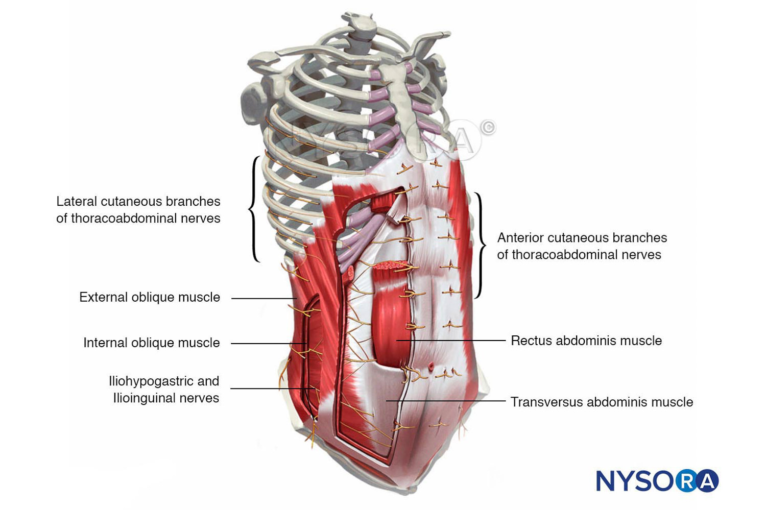
Abdominal wall muscles and innervation. The anatomy of the muscles of the abdominal wall can be seen in this image. Also the nerves that innervate these muscles and their location are shown. English labels.
Image created for NYSORA by VisionExpo.Design - www.nysora.com
Image created for NYSORA by VisionExpo.Design - www.nysora.com
Anatomical structures in item:
Uploaded by: rva
Netherlands, Leiden – Leiden University Medical Center, Leiden University
Cavea thoracis
Musculus obliquus externus abdominis
Musculus obliquus internus abdominis
Nervus iliohypogastricus
Nervus ilioinguinalis
Musculus rectus abdominis
Musculus transversus abdominis
Creator(s)/credit: New York School of Regional Anesthesia; VisionExpo.Design, illustration creation
Requirements for usage
You are free to use this item if you follow the requirements of the license:  View license
 View license
 If you use this item you should credit it as follows:
- For usage in print - copy and paste the line below:
- For digital usage (e.g. in PowerPoint, Impress, Word, Writer) - copy and paste the line below (optionally add the license icon):
"NYSORA - Drawing Abdominal wall muscles and innervation - English labels" at AnatomyTOOL.org by New York School of Regional Anesthesia and VisionExpo.Design, license: Creative Commons Attribution-NonCommercial-NoDerivs


Comments