
nid: 62836
Additional formats:
None available
Description:
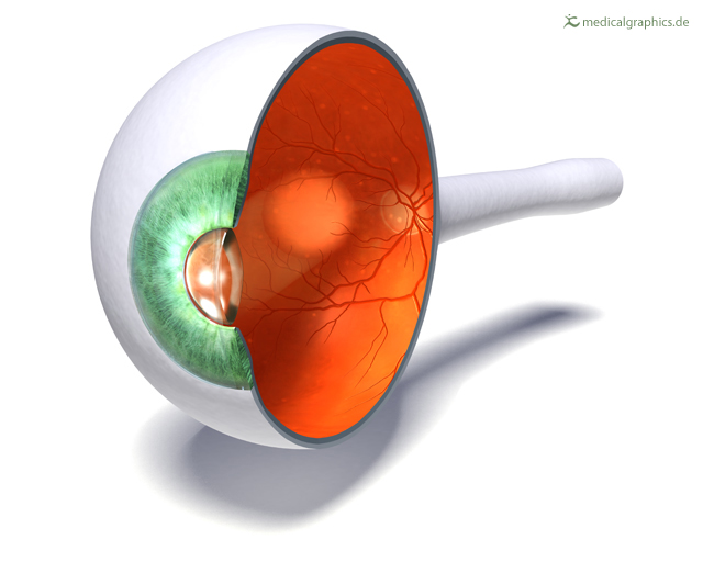
Eye sagittal section. The cornea and retina can be seen because of the cross-section.
Retrieved from www.MedicalGraphics.de. Similar images, including pathology can be found here.
Retrieved from www.MedicalGraphics.de. Similar images, including pathology can be found here.
Anatomical structures in item:
Uploaded by: rva
Netherlands, Leiden – Leiden University Medical Center, Leiden University
Oculus
Retina
Oculus et structurae pertinentes
Nervus opticus
Creator(s)/credit: www.MedicalGraphics.de
Requirements for usage
You are free to use this item if you follow the requirements of the license:  View license
View license If you use this item you should credit it as follows:
- For usage in print - copy and paste the line below:
- For digital usage (e.g. in PowerPoint, Impress, Word, Writer) - copy and paste the line below (optionally add the license icon):
"MedicalGraphics - Drawing Eye sagittal section - no labels" at AnatomyTOOL.org by www.MedicalGraphics.de, license: Creative Commons Attribution-NoDerivatives


Comments