
nid: 60082
Additional formats:
None available
Description:
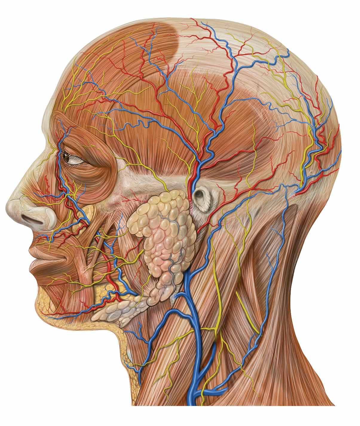
Superficial anatomy of the head from lateral. In this image, several superficial structures of the face and neck can be seen, including facial muscles, the parotid gland, nerves and blood vessels. No labels
Anatomical structures in item:
Uploaded by: rva
Netherlands, Leiden – Leiden University Medical Center, Leiden University
Cranium
Glandula parotidea
Venter frontalis musculus occipitofrontalis
Musculus orbicularis oris
Musculus orbicularis oculi
Musculus nasalis
Oculus
Meatus acusticus externus
Arcus zygomaticus
Musculus sternocleidomastoideus
Creator(s)/credit: Patrick J. Lynch, medical illustrator; C. Carl Jaffe MD, cardiologist
Requirements for usage
You are free to use this item if you follow the requirements of the license:  View license
View license If you use this item you should credit it as follows:
- For usage in print - copy and paste the line below:
- For digital usage (e.g. in PowerPoint, Impress, Word, Writer) - copy and paste the line below (optionally add the license icon):
"Lynch - Drawing Superficial anatomy of the head from lateral - no labels" at AnatomyTOOL.org by Patrick J. Lynch and C. Carl Jaffe, license: Creative Commons Attribution


Comments