
nid: 62217
Additional formats:
None available
Description:
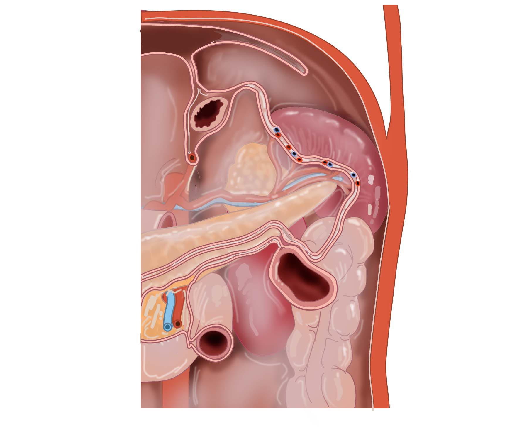
This image gives an impression of the varying numbers of original peritoneal layers at different locations. These layers later often adhere to each other where they lie against each other. Drawings often show the postnatal situation depicting only the resulting single peritoneal layer. This makes it very difficult to visualize how the peritoneum has developed. To understand, one must visualize the original embryological situation with its several peritoneal layers, as shown in this drawing.
Anatomical structures in item:
Uploaded by: rva
Netherlands, Leiden – Leiden University Medical Center, Leiden University
Abdomen
Spatium retroperitoneale
Splen
Colon descendens
Duodenum
Pancreas
Cauda pancreatis
Arteria lienalis
Vena lienalis
Ren (Nephros)
Flexura coli sinistra
Ligamentum splenocolicum
Oesophagus
Creator(s)/credit: Ron Slagter NZIMBI, medical illustrator; O. Paul Gobée MD, anatomist, LUMC
Requirements for usage
You are free to use this item if you follow the requirements of the license:  View license
View license If you use this item you should credit it as follows:
- For usage in print - copy and paste the line below:
- For digital usage (e.g. in PowerPoint, Impress, Word, Writer) - copy and paste the line below (optionally add the license icon):
"Leiden - Drawing Varying numbers of original peritoneal layers per location - no labels" at AnatomyTOOL.org by Ron Slagter and O. Paul Gobée, LUMC, license: Creative Commons Attribution-NonCommercial-ShareAlike


Comments