

nid: 62811
Additional formats:
None available
Description:
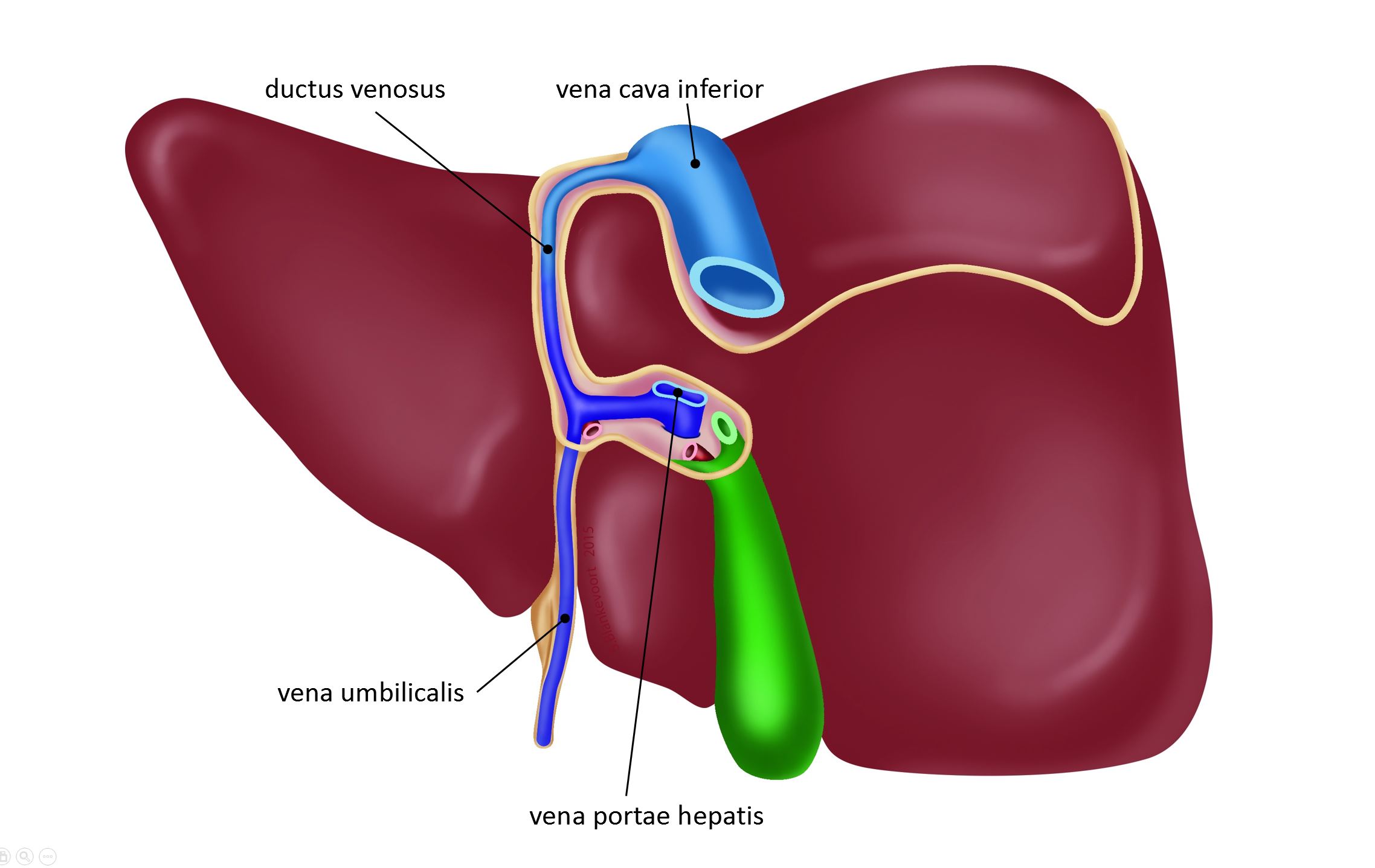
Posterior view of the prenatal liver. Different from the postnatal posterior view are the presence of the ductus venosus (postnatal: ligamentum venosum) and the umbilical vein (postnatal: round ligament).
Labels added by Paul Gobée.
Labels added by Paul Gobée.
Anatomical structures in item:
Uploaded by: admin
Netherlands, Leiden – Leiden University Medical Center, Leiden University
Hepar
Corpus vesicae biliaris
Fossa vesicae biliaris
Vena cava inferior
Vena portae hepatis
Arteria hepatica propria
Ductus biliaris
Ligamentum venosum
Vena umbilicalis
Creator(s)/credit: Bas Blankevoort, medical illustrator, LUMC; O.Paul Gobée MD, anatomist, LUMC
Requirements for usage
You are free to use this item if you follow the requirements of the license:  View license
View license If you use this item you should credit it as follows:
- For usage in print - copy and paste the line below:
- For digital usage (e.g. in PowerPoint, Impress, Word, Writer) - copy and paste the line below (optionally add the license icon):
"Leiden - Drawing Posterior view prenatal liver with ductus venosus - Latin labels" at AnatomyTOOL.org by Bas Blankevoort, LUMC and O.Paul Gobée, LUMC, license: Creative Commons Attribution-NonCommercial-ShareAlike
"Leiden - Drawing Posterior view prenatal liver with ductus venosus - Latin labels" by Bas Blankevoort, LUMC and O.Paul Gobée, LUMC, license: CC BY-NC-SA


Comments