
nid: 62382
Additional formats:
None available
Description:
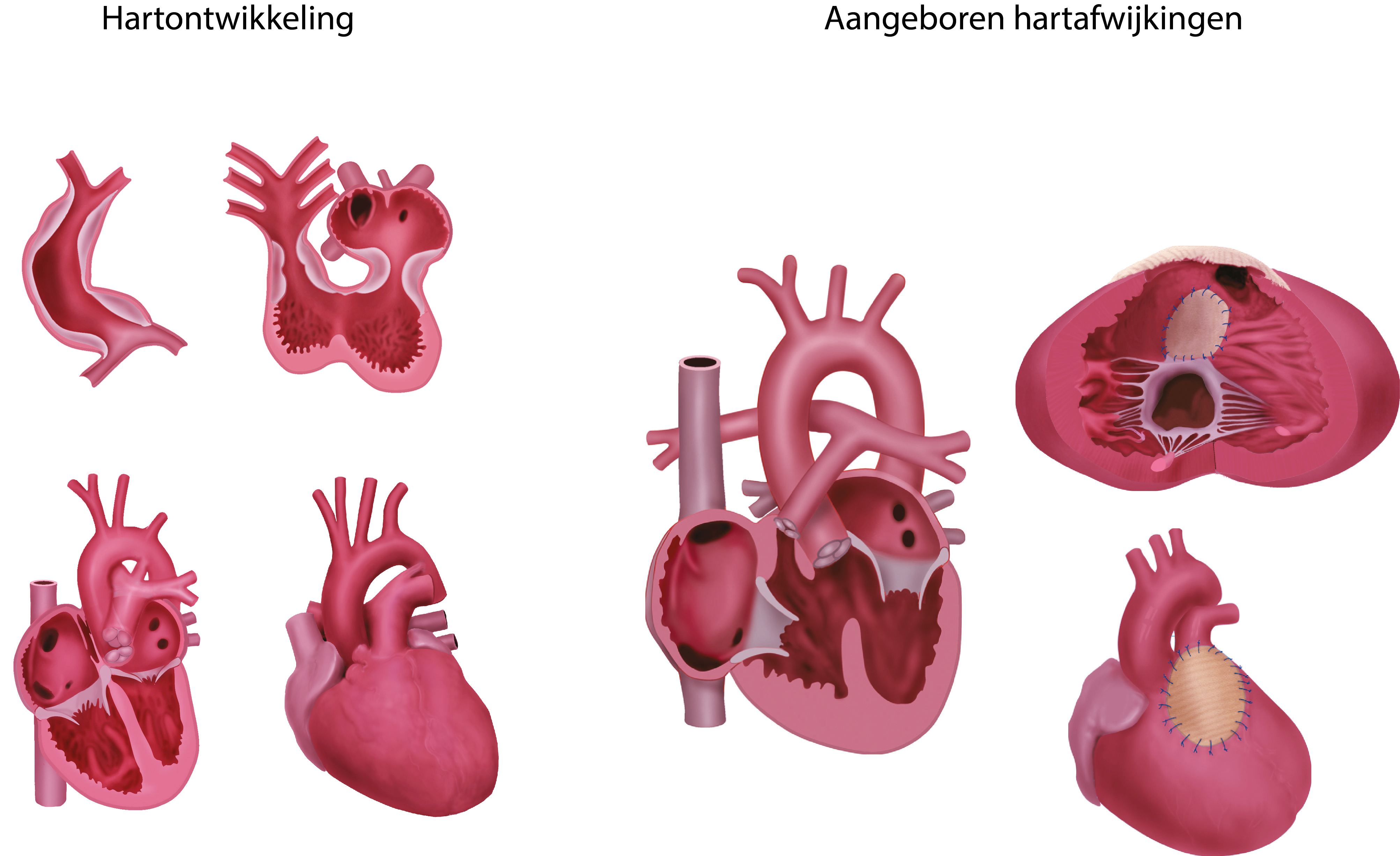
Normal heart development and tetralogy of Fallot. In the four left drawings, the embryonal heart development is shown and in the three right drawings, the congenital heart disease tetralogy of Fallot is shown (before (a) and after (b) surgery). Version without labels
Anatomical structures in item:
Uploaded by: rva
Netherlands, Leiden – Leiden University Medical Center, Leiden University
Cor
Endocardium
Vena cava superior
Atrium dextrum
Atrium sinistrum
Ventriculus sinister
Ventriculus dexter
Vena cava inferior
Aorta
Truncus pulmonalis
Creator(s)/credit: Ron Slagter NZIMBI, medical illustrator; prof Adri C. Gittenberger-de Groot PhD, anatomist, head of dept. anatomy & embryology, LUMC; Dr Monique R.M. Jongbloed PhD, cardiologist, anatomist, LUMC; Dr. Margot M. Bartelings MD, PhD, anatomist, LUMC
Requirements for usage
You are free to use this item if you follow the requirements of the license:  View license
View license If you use this item you should credit it as follows:
- For usage in print - copy and paste the line below:
- For digital usage (e.g. in PowerPoint, Impress, Word, Writer) - copy and paste the line below (optionally add the license icon):
"Leiden - Drawing Normal heart development and tetralogy of Fallot - no labels" at AnatomyTOOL.org by Ron Slagter, Adri C. Gittenberger-de Groot, LUMC, Monique R.M. Jongbloed, LUMC et al, license: Creative Commons Attribution-NonCommercial-ShareAlike


Comments