
nid: 62110
Additional formats:
None available
Description:
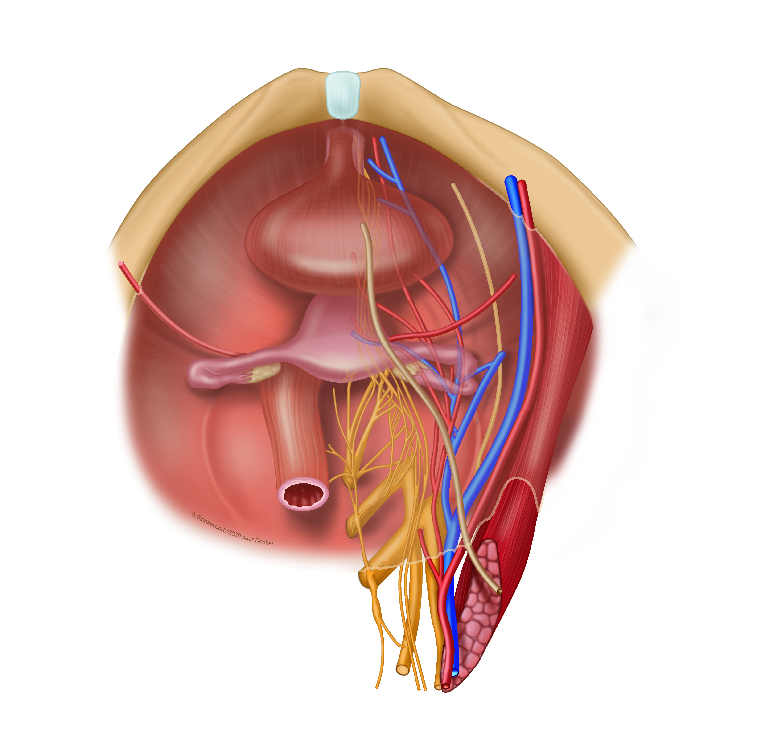
Female pelvic floor organs. The pelvic floor (diaphragm) is the lowest part of the pelvic cavity.
Coloured illustration based on the black-white drawings of prof dr P.J. Donker in 1982 (LUMC).
Coloured illustration based on the black-white drawings of prof dr P.J. Donker in 1982 (LUMC).
Anatomical structures in item:
Uploaded by: rva
Netherlands, Leiden – Leiden University Medical Center, Leiden University
Diaphragma pelvis
Symphysis pubica
Uterus
Tuba uterina (Salpinx)
Ovarium
Vesica urinaria
Ureter
Creator(s)/credit: Bas (S) Blankevoort, biology and medical illustrator, LUMC; Prof. Marco C DeRuiter PhD, anatomist, professor of clinical and applied anatomy, LUMC
Requirements for usage
You are free to use this item if you follow the requirements of the license:  View license
View license If you use this item you should credit it as follows:
- For usage in print - copy and paste the line below:
- For digital usage (e.g. in PowerPoint, Impress, Word, Writer) - copy and paste the line below (optionally add the license icon):
"Leiden - Drawing Female pelvic floor organs - no labels" at AnatomyTOOL.org by Bas (S) Blankevoort, LUMC and Marco C DeRuiter, LUMC, license: Creative Commons Attribution-NonCommercial-ShareAlike
"Leiden - Drawing Female pelvic floor organs - no labels" by Bas (S) Blankevoort, LUMC and Marco C DeRuiter, LUMC, license: CC BY-NC-SA


Comments