
nid: 59148
Additional formats:
None available
Description:
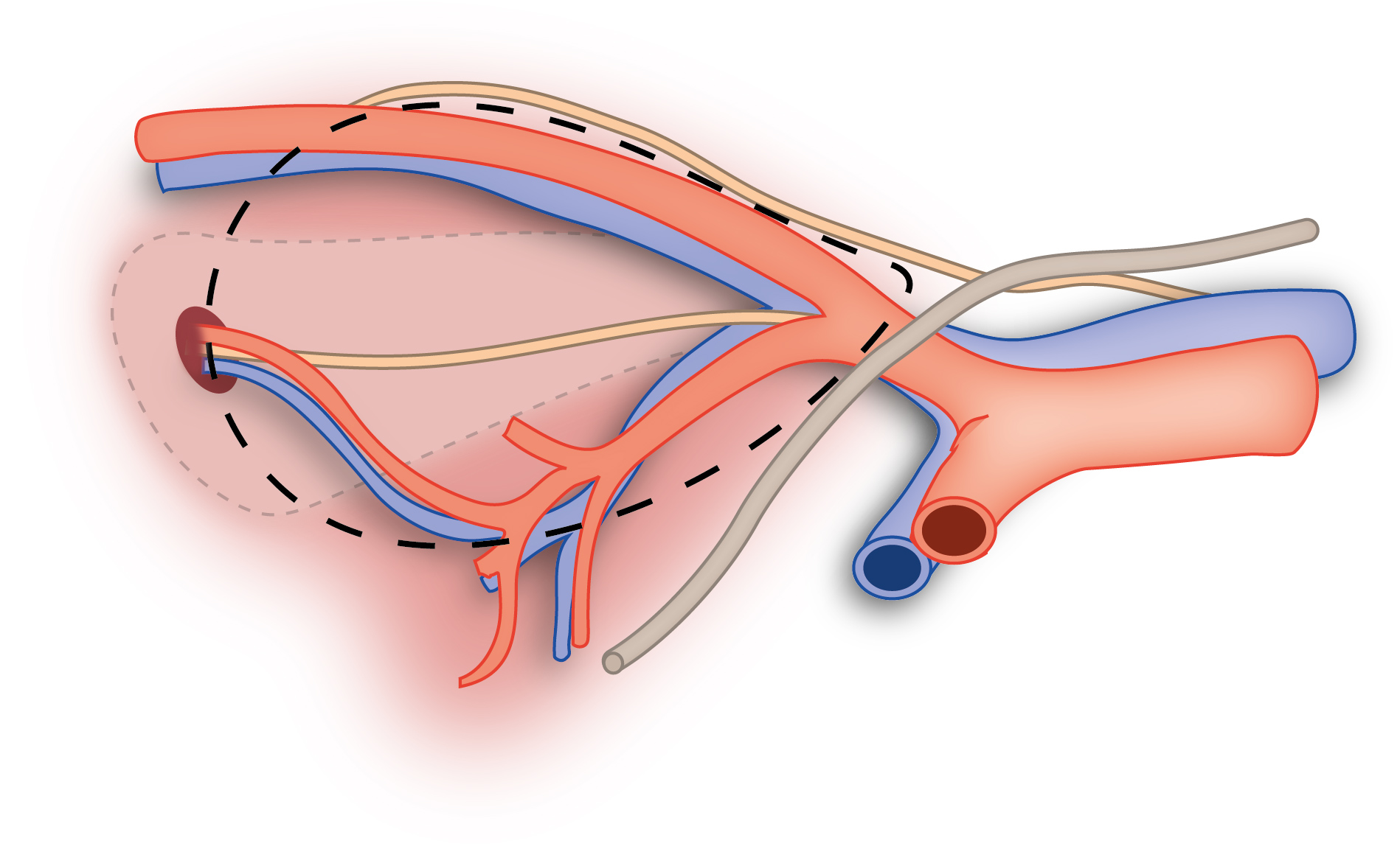
Lateral view of area of lymph node dissection in bladder cancer. Dotted line outlines the area of dissection. No labels.
Illustration by Ron Slagter and Marco DeRuiter for course 'Surgical Anatomy of the lesser pelvis' by the 'Urologisch Opleidings Instituut', the Netherlands.
Illustration by Ron Slagter and Marco DeRuiter for course 'Surgical Anatomy of the lesser pelvis' by the 'Urologisch Opleidings Instituut', the Netherlands.
Anatomical structures in item:
Uploaded by: Siem Zethof
Netherlands, Leiden – Leiden University Medical Center, Leiden University
Bifurcatio aortae
Ureter
Arteria iliaca externa
Arteria iliaca interna
Arteria obturatoria
Nervus obturatorius
Creator(s)/credit: Ron Slagter NZIMBI, medical illustrator, LUMC; Prof. Marco DeRuiter PhD, anatomist, LUMC
Requirements for usage
You are free to use this item if you follow the requirements of the license:  View license
View license If you use this item you should credit it as follows:
- For usage in print - copy and paste the line below:
- For digital usage (e.g. in PowerPoint, Impress, Word, Writer) - copy and paste the line below (optionally add the license icon):
"Lateral view of area of lymph node dissection in bladder cancer – no labels" at AnatomyTOOL.org by Ron Slagter, LUMC and Marco DeRuiter, LUMC, license: Creative Commons Attribution-NonCommercial-ShareAlike
"Lateral view of area of lymph node dissection in bladder cancer – no labels" by Ron Slagter, LUMC and Marco DeRuiter, LUMC, license: CC BY-NC-SA


Comments