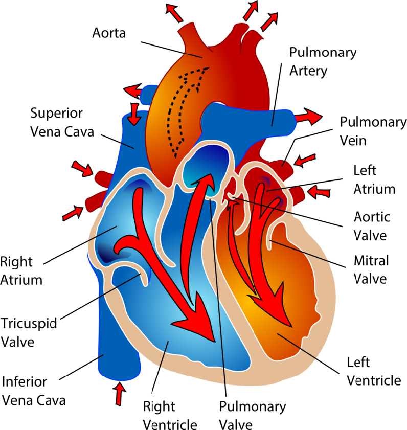
Human Biology fig. 1.54 - Bloodflow in the heart - English labels

nid: 59796
Additional formats:
None available
Description:
Bloodflow in the heart. The chambers of the heart and the valves between them are shown in this image, moreover the bloodflow is labeled with arrows. English labels. 

From ‘Human Biology’ by D. Wilkin and J. Brainard.

 
Anatomical structures in item:
Aorta
Vena cava superior
Atrium dextrum
Valva tricuspidalis
Vena cava inferior
Ventriculus dexter
Valva trunci pulmonalis
Ventriculus sinister
Truncus pulmonalis
Atrium sinistrum
Venae pulmonales
Valva aortae
Valva mitralis
Ventriculus sinister
Cor
Uploaded by: rva Netherlands, Leiden – Leiden University Medical Center, Leiden University
Creator(s)/credit: Mariana Ruiz Villarreal (Wikimedia: LadyofHats)
Requirements for usage
You are free to use this item. Read more

This item is in the Public Domain because its copyright has expired. You are not required to credit its creators when you use it. Nevertheless, it is adviced to do so. First, it is academically correct to pay tribute to the creators. Second, items of unknown origin might be classified as 'copyright infringement' by copyright controlling bodies, with possible resulting bills. Stating the item's source will prevent this. You can use the following text:

  • For usage in print - copy and paste the line below:
  • "Human Biology fig. 1.54 - Bloodflow in the heart - English labels" at AnatomyTOOL.org by Mariana Ruiz Villarreal (Wikimedia: LadyofHats) is in the Public Domain. Modified by CK-12 Foundation. Source: book ‘Human Biology’, https://textbookequity.org/Textbooks/HumanBiologyCK12.pdf..
  • For digital usage (e.g. in PowerPoint, Impress, Word, Writer) - copy and paste the line below (optionally add the icon):
  • "Human Biology fig. 1.54 - Bloodflow in the heart - English labels" by Mariana Ruiz Villarreal (Wikimedia: LadyofHats) is in the Public Domain.. Modified by CK-12 Foundation. Source: book ‘Human Biology’, https://textbookequity.org/Textbooks/HumanBiologyCK12.pdf.

254 reads
Human Biology fig. 1.54 - Bloodflow in the heart - English labels
Uploaded by: rva
Institution: Netherlands, Leiden – Leiden University Medical Center, Leiden University
Creator(s)/credit: Mariana Ruiz Villarreal (Wikimedia: LadyofHats)
Review
  • Status: to be reviewed
Bloodflow in the heart. The chambers of the heart and the valves between them are shown in this image, moreover the bloodflow is labeled with arrows. English labels. 

From ‘Human Biology’ by D. Wilkin and J. Brainard.

 
User comments
Language
EN
Educational level
+ + +
Technical info
Item id ('nid'): 59796
*.jpg, 190kB, 796x841
User comments

Comments

Anatomical structures in item
Aorta
Vena cava superior
Atrium dextrum
Valva tricuspidalis
Vena cava inferior
Ventriculus dexter
Valva trunci pulmonalis
Ventriculus sinister
Truncus pulmonalis
Atrium sinistrum
Venae pulmonales
Valva aortae
Valva mitralis
Ventriculus sinister
Cor
Topics
Gross
Image / technique source
Drawing