
nid: 60529
Additional formats:
None available
Description:
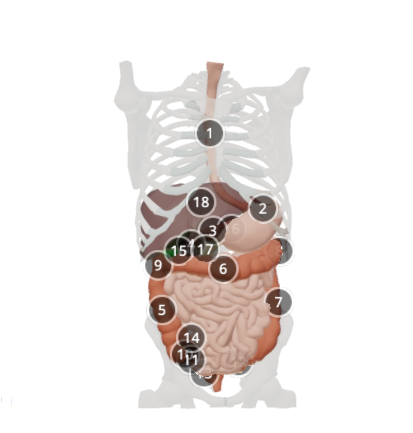
This model depicts important organs in the gastrointestinal tract. Data from CT scan was transformed into 3D objects in Invesalius, imported to Zbrush 2018 and further modified, based on specimen in the dissection room, CT data of this particular patient and references in anatomy books.
Anatomical structures in item:
Uploaded by: rva
Netherlands, Leiden – Leiden University Medical Center, Leiden University
Oesophagus
Ventriculus
Pylorus
Duodenum
Colon ascendens
Colon transversum
Colon descendens
Flexura coli sinistra
Flexura coli dextra
Caecum
Appendix vermiformis
Colon sigmoideum
Rectum
Ileocecal junction
Vesica biliaris (Fellea)
Corpus pancreatis
Caput pancreatis
Hepar
Creator(s)/credit: Anna Sieben BM, MSc, medical artist and scientific illustrator, UMCG; Cyril Luman MSc, UMCG; Walter Noordzij MD, Nuclear Medicine Physician, UMCG
Requirements for usage
You are free to use this item if you follow the requirements of the license:  View license
View license If you use this item you should credit it as follows:
- For usage in print - copy and paste the line below:
- For digital usage (e.g. in PowerPoint, Impress, Word, Writer) - copy and paste the line below (optionally add the license icon):
"Groningen - 3D model Gastrointestinal tract" at AnatomyTOOL.org by Anna Sieben, UMCG, Cyril Luman, UMCG and Walter Noordzij, UMCG, license: Creative Commons Attribution-NonCommercial-ShareAlike
"Groningen - 3D model Gastrointestinal tract" by Anna Sieben, UMCG, Cyril Luman, UMCG and Walter Noordzij, UMCG, license: CC BY-NC-SA


Comments