
nid: 60459
Additional formats:
None available
Description:
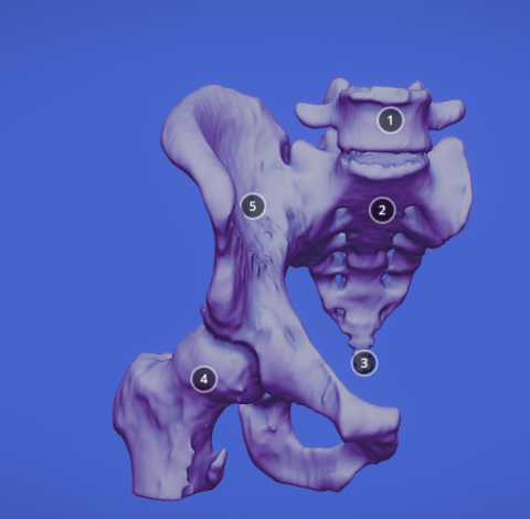
In this 3D model, the main bones of the male pelvis are shown. The left iliac bone has been separated to show the sacroiliac joint.
Anatomical structures in item:
Uploaded by: rva
Netherlands, Leiden – Leiden University Medical Center, Leiden University
Pelvis
Vertebrae lumbales (LI-LV)
Os coxae
Ilium
Femur
Os sacrum [vertebrae sacrales I - V]
Creator(s)/credit: Ramon Gonzalez Cabrera
Requirements for usage
You are free to use this item if you follow the requirements of the license:  View license
View license If you use this item you should credit it as follows:
- For usage in print - copy and paste the line below:
- For digital usage (e.g. in PowerPoint, Impress, Word, Writer) - copy and paste the line below (optionally add the license icon):
"Gonzalez - 3D model Male Pelvis" at AnatomyTOOL.org by Ramon Gonzalez Cabrera, license: Creative Commons Attribution
"Gonzalez - 3D model Male Pelvis" by Ramon Gonzalez Cabrera, license: CC BY


Comments