
nid: 57776
Additional formats:
None available
Description:
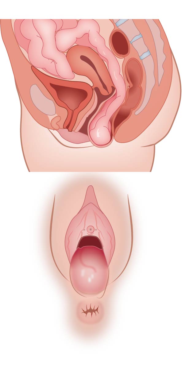
Enterocele on saggital view. At vaginal examination a posterior vaginal wall prolapse can be seen - no labels
Illustration by Ron Slagter.
Illustration by Ron Slagter.
Anatomical structures in item:
Uploaded by: opgobee
Netherlands, Leiden – Leiden University Medical Center, Leiden University
Pelvis
Rectum
Creator(s)/credit: Ron Slagter NZIMBI, medical illustrator, LUMC
Requirements for usage
You are free to use this item if you follow the requirements of the license:  View license
View license If you use this item you should credit it as follows:
- For usage in print - copy and paste the line below:
- For digital usage (e.g. in PowerPoint, Impress, Word, Writer) - copy and paste the line below (optionally add the license icon):
"Enterocele - no labels" at AnatomyTOOL.org by Ron Slagter, LUMC, license: Creative Commons Attribution-NonCommercial-ShareAlike
"Enterocele - no labels" by Ron Slagter, LUMC, license: CC BY-NC-SA


Comments