
nid: 60464
Additional formats:
None available
Description:
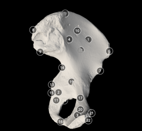
This 3D model shows the anatomy of the left pelvic bone with annotations.
Anatomical structures in item:
Uploaded by: rva
Netherlands, Leiden – Leiden University Medical Center, Leiden University
Os coxae
Pelvis
Ilium
Ischium
Pubis
Ala ossis ilii
Crista iliaca
Spina iliaca anterior superior
Spina iliaca anterior inferior
Spina iliaca posterior superior
Spina iliaca posterior inferior
Fossa iliaca
Ramusi ossis ischii
Foramen obturatum
Spina ischiadica
Incisura ischiadica major
Incisura ischiadica minor
Ramus inferior ossis pubis
Ramus superior ossis pubis
Tuber ischiadicum
Acetabulum
Corpus ossis pubis
Crista pubica
Tuberculum pubicum
Pelvis major
Creator(s)/credit: Dr Eric Bauer, Biology professor
Requirements for usage
You are free to use this item if you follow the requirements of the license:  View license
View license If you use this item you should credit it as follows:
- For usage in print - copy and paste the line below:
- For digital usage (e.g. in PowerPoint, Impress, Word, Writer) - copy and paste the line below (optionally add the license icon):
"Elon - 3D model Pelvic bone" at AnatomyTOOL.org by Eric Bauer, license: Creative Commons Attribution. Scanned by: students in Dr. Eric Bauer’s human anatomy lab at Elon University, North Carolina, USA.
"Elon - 3D model Pelvic bone" by Eric Bauer, license: CC BY. Scanned by: students in Dr. Eric Bauer’s human anatomy lab at Elon University, North Carolina, USA.


Comments