
nid: 59842
Additional formats:
None available
Description:
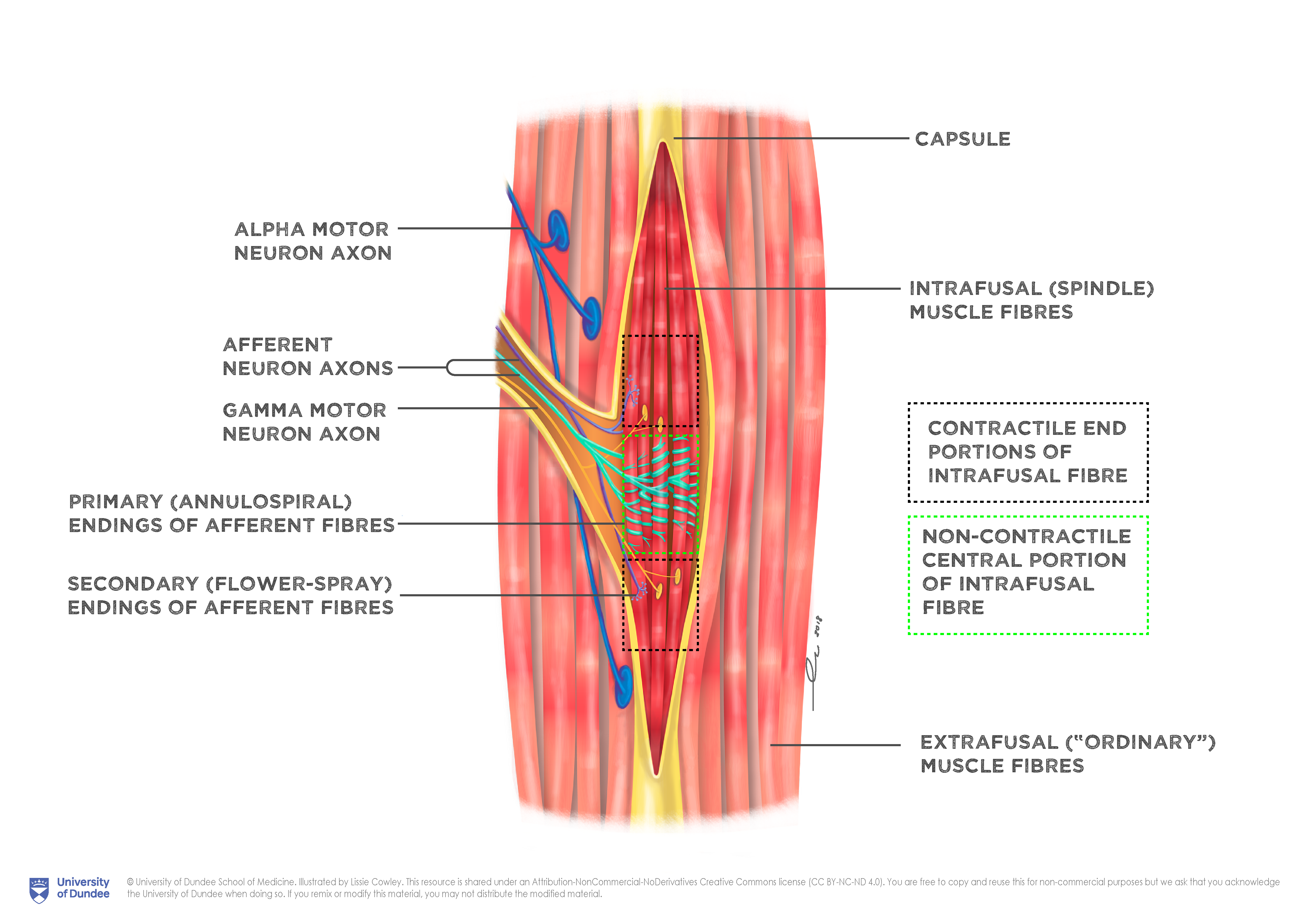
Anatomy of the muscle spindle with callout boxes. Muscle spindles are stretch receptors within muscles that primarily detect changes in the length of the muscle. The boxes lined with black dots depict the contractile end portions of intrafusal fibre while the box lined with green dots depicts the non-contractile central portion of the intrafusal fibre. English labels. NOTE: THIS IMAGE IS UNDER A NON-DERIVATIVE LICENSE. THIS MEANS THAT IF YOU REMIX OR REVISE THIS MATERIAL YOU MAY NOT DISTRIBUTE THE MODIFIED MATERIAL.
Anatomical structures in item:
Uploaded by: rva
Netherlands, Leiden – Leiden University Medical Center, Leiden University
Neurofibrae afferentes
Neurofibrae efferentes
Creator(s)/credit: Lissie Cowley MSc, medical illustrator
Requirements for usage
You are free to use this item if you follow the requirements of the license:  View license
View license If you use this item you should credit it as follows:
- For usage in print - copy and paste the line below:
- For digital usage (e.g. in PowerPoint, Impress, Word, Writer) - copy and paste the line below (optionally add the license icon):
"Dundee - Drawing Anatomy of the muscle spindle with callout boxes - English labels" at AnatomyTOOL.org by Lissie Cowley, © University of Dundee School of Medicine, license: Creative Commons Attribution-NonCommercial-NoDerivs
"Dundee - Drawing Anatomy of the muscle spindle with callout boxes - English labels" by Lissie Cowley, © University of Dundee School of Medicine, license: CC BY-NC-ND


Comments