nid: 59550
Additional formats:
None available
Description:
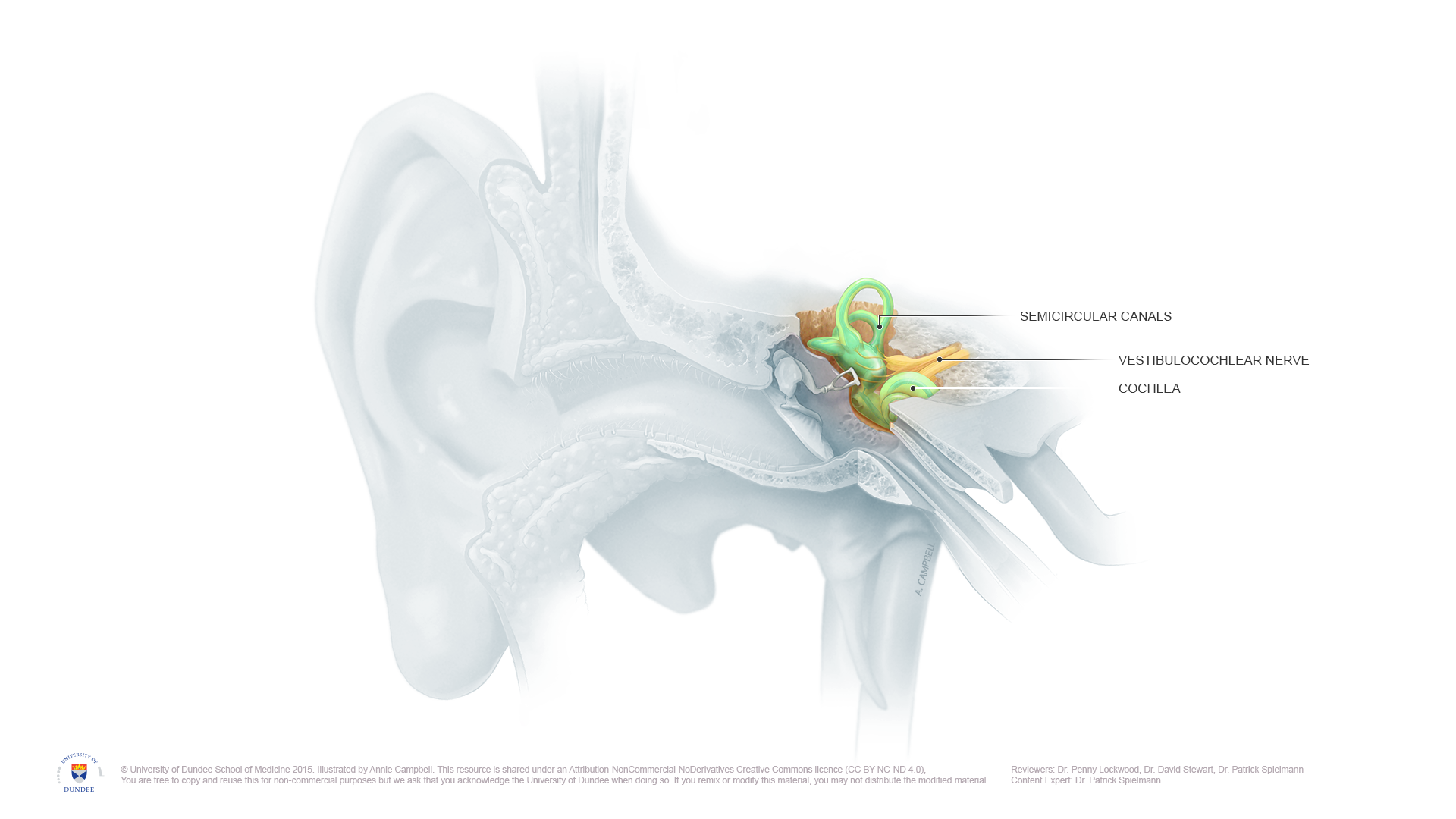
Anatomy of Inner Ear. The anatomy of the inner ear can be appreciated. English labels.
NOTE: THIS IMAGE IS UNDER A NON-DERIVATIVE LICENSE. THIS MEANS THAT IF YOU REMIX OR REVISE THIS MATERIAL YOU MAY NOT DISTRIBUTE THE MODIFIED MATERIAL.
NOTE: THIS IMAGE IS UNDER A NON-DERIVATIVE LICENSE. THIS MEANS THAT IF YOU REMIX OR REVISE THIS MATERIAL YOU MAY NOT DISTRIBUTE THE MODIFIED MATERIAL.
Anatomical structures in item:
Uploaded by: rva
Netherlands, Leiden – Leiden University Medical Center, Leiden University
Auris interna
Canales semicirculares
Nervus vestibulocochlearis [VIII]
Cochlea
Creator(s)/credit: Annie Campbell MSc, medical illustrator
Requirements for usage
You are free to use this item if you follow the requirements of the license:  View license
 View license
 If you use this item you should credit it as follows:
- For usage in print - copy and paste the line below:
- For digital usage (e.g. in PowerPoint, Impress, Word, Writer) - copy and paste the line below (optionally add the license icon):
"Dundee - Drawing Anatomy of Inner Ear - English labels" at AnatomyTOOL.org by Annie Campbell, © University of Dundee School of Medicine, license: Creative Commons Attribution-NonCommercial-NoDerivs. Reviewed by: Dr. Penny Lockwood, Dr. David Stewart, Dr. Patrick Spielmann
"Dundee - Drawing Anatomy of Inner Ear - English labels" by Annie Campbell, © University of Dundee School of Medicine, license: CC BY-NC-ND. Reviewed by: Dr. Penny Lockwood, Dr. David Stewart, Dr. Patrick Spielmann


Comments