nid: 60505
Additional formats:
None available
Description:
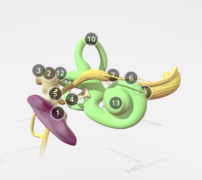
This model shows the anatomy of the inner ear. Certain aspects of this model were created from segmented MRI data, making this a highly accurate representation of the tympanic membrane, facial nerve, ossicles and vestibular system.
Anatomical structures in item:
Uploaded by: rva
Netherlands, Leiden – Leiden University Medical Center, Leiden University
Membrana tympanica
Malleus
Incus
Stapes
Chorda tympani
Nervus petrosus major
Nervus facialis [VII]
Nervus vestibulocochlearis [VIII]
Nervus cochlearis
Nervus vestibularis
Canalis semicircularis anterior
Canalis semicircularis posterior
Canalis semicircularis lateralis
Cochlea
Auris interna
Auris
Creator(s)/credit: Annie Campbell, Dundee; University of Dundee School of Medicine; Prof W. Robert J. Funnel PhD, McGill
Requirements for usage
You are free to use this item if you follow the requirements of the license:  View license
 View license
 If you use this item you should credit it as follows:
- For usage in print - copy and paste the line below:
- For digital usage (e.g. in PowerPoint, Impress, Word, Writer) - copy and paste the line below (optionally add the license icon):
"Dundee - 3D model Anatomy of the Inner Ear - numbered English labels" at AnatomyTOOL.org by Annie Campbell, Dundee, University of Dundee School of Medicine and W. Robert J. Funnel, McGill, license: Creative Commons Attribution-NonCommercial-ShareAlike. Derivative of: “3D Ear” by W. Robert J. Funnell, PhD; Sam Daniel, MD, CM; and Daren Nicolson, MD, CM at McGill University
"Dundee - 3D model Anatomy of the Inner Ear - numbered English labels" by Annie Campbell, Dundee, University of Dundee School of Medicine and W. Robert J. Funnel, McGill, license: CC BY-NC-SA. Derivative of: “3D Ear” by W. Robert J. Funnell, PhD; Sam Daniel, MD, CM; and Daren Nicolson, MD, CM at McGill University


Comments