
nid: 62591
Additional formats:
None available
Description:
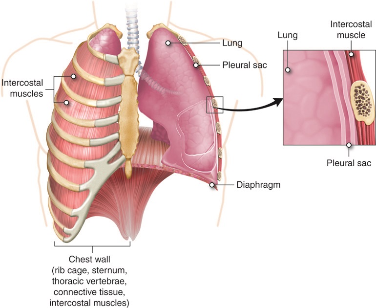
Lungs and chest wall. The chest wall surrounds the organs in the thorax. The wall consists of the rib cage, sternum, thoracic vertebrae, connective tissue and intervoastal muscles. On the left side, the rib cage is removed. English labels.
Retrieved from Anatomy & Physiology by Open Learning Initiative (CC BY-NC-SA).
Retrieved from Anatomy & Physiology by Open Learning Initiative (CC BY-NC-SA).
Anatomical structures in item:
Uploaded by: rva
Netherlands, Leiden – Leiden University Medical Center, Leiden University
Pulmones
Musculus intercostalis
Thorax
Sternum
Cavea thoracis
Diaphragma
Os costale (Costa)
Creator(s)/credit: Cenveo
Requirements for usage
You are free to use this item if you follow the requirements of the license:  View license
View license If you use this item you should credit it as follows:
- For usage in print - copy and paste the line below:
- For digital usage (e.g. in PowerPoint, Impress, Word, Writer) - copy and paste the line below (optionally add the license icon):
"Cenveo - Drawing Lungs and chest wall - English labels" at AnatomyTOOL.org by Cenveo, license: Creative Commons Attribution
"Cenveo - Drawing Lungs and chest wall - English labels" by Cenveo, license: CC BY


Comments