nid: 59515
Additional formats:
None available
Description:
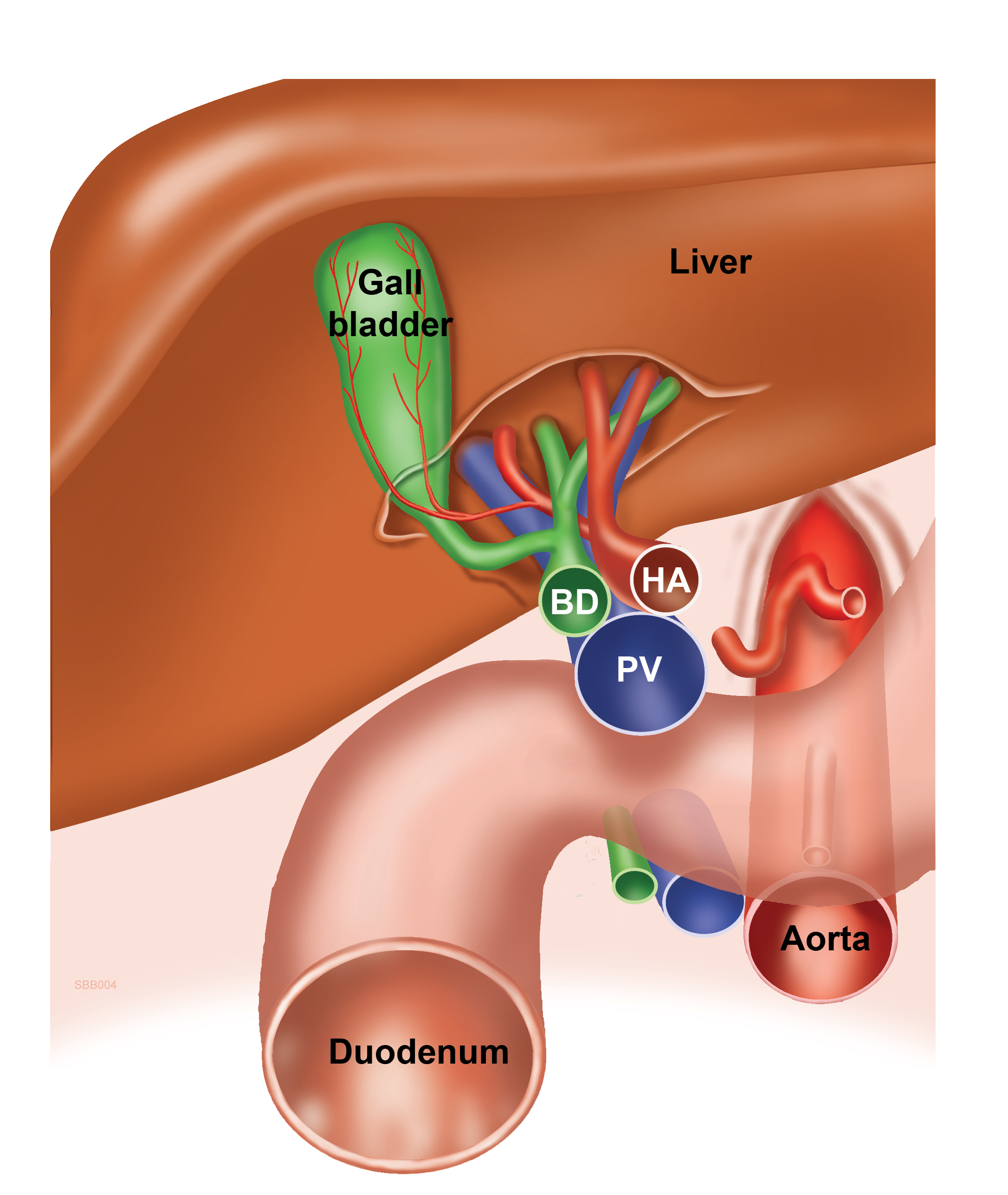
This image schematically shows the positioning of the three structures in the hepatoduodenal ligament: the hepatic artery lies ventromedially, the bile duct lateromedially and the portal vein dorsally. Their positions can be memorized by visualizing how the shown cross-section (a larger circle with two smaller circles on top) resembles a well known cartoon mouse. The large circle below that resembles the mouse's head is the portal vein, the smaller circle on the top medial side that resembles an ear is the (proper) hepatic artery and the smaller circle on the top lateral side that resembles the other ear is the bile duct. The idea for this presentation was given by prof. Andrzej Baranski, surgeon at Leiden University Medical Center (LUMC), the Netherlands. The image was created at the dept. of Anatomy and Embryology of LUMC.
Anatomical structures in item:
Uploaded by: opgobee
Netherlands, Leiden – Leiden University Medical Center, Leiden University
Ligamentum hepatoduodenale
Vena portae hepatis
Arteria hepatica propria
Ductus biliaris
Hepar
Creator(s)/credit: S. Bas Blankevoort, medical illustrator, LUMC; Prof. Andrzej Baranski MD, PhD, surgeon, LUMC; O. Paul Gobée MD, anatomist and e-learning developer, LUMC
Requirements for usage
You are free to use this item if you follow the requirements of the license:  View license
 View license
 If you use this item you should credit it as follows:
- For usage in print - copy and paste the line below:
- For digital usage (e.g. in PowerPoint, Impress, Word, Writer) - copy and paste the line below (optionally add the license icon):
"Cartoon to remember the position of the structures in the hepatoduodenal ligament - English labels" at AnatomyTOOL.org by S. Bas Blankevoort, LUMC, Andrzej Baranski, LUMC and O. Paul Gobée, LUMC, license: Creative Commons Attribution-NonCommercial-ShareAlike
"Cartoon to remember the position of the structures in the hepatoduodenal ligament - English labels" by S. Bas Blankevoort, LUMC, Andrzej Baranski, LUMC and O. Paul Gobée, LUMC, license: CC BY-NC-SA


Comments