
nid: 58602
Additional formats:
None available
Description:
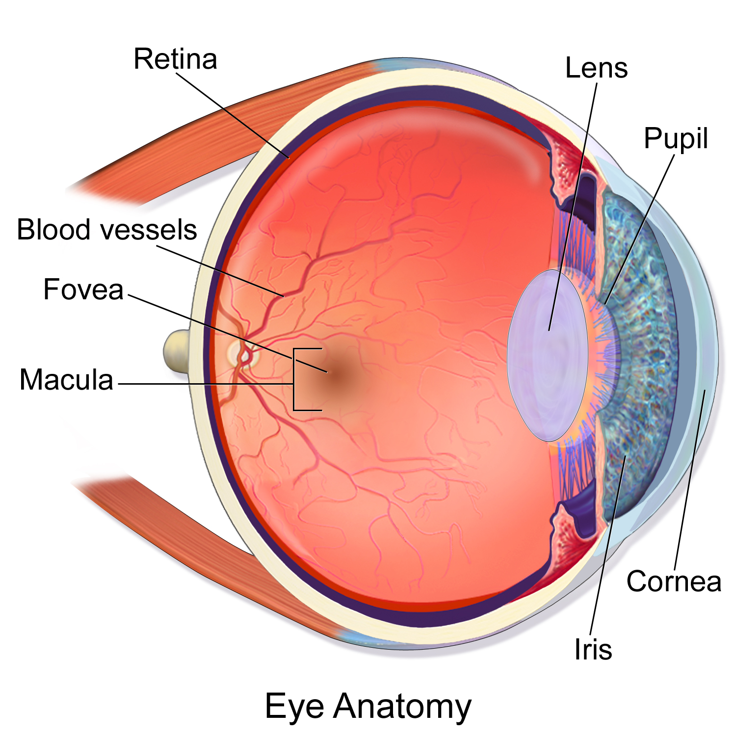
Anatomy of the eye. Sagittal section of the eye showing the anatomy. English labels.
By Blausen.com staff (2014). Retrieved from Medical gallery of Blausen Medical 2014., WikiJournal of Medicine 1 (2): 10. DOI:10.15347/wjm/2014.010. ISSN 2002-4436. Fig. 0389.
By Blausen.com staff (2014). Retrieved from Medical gallery of Blausen Medical 2014., WikiJournal of Medicine 1 (2): 10. DOI:10.15347/wjm/2014.010. ISSN 2002-4436. Fig. 0389.
Anatomical structures in item:
Uploaded by: Student10
Netherlands, Leiden – Leiden University Medical Center, Leiden University
Oculus
Cornea
Retina
Choroidea
Sclera
Iris
Lens
Camera vitrea bulbi oculi
Discus nervi optici
Fovea centralis
Macula lutea
Pupilla
Musculus rectus superior
Musculus rectus inferior
Creator(s)/credit: Blausen.com staff (2014)
Requirements for usage
You are free to use this item if you follow the requirements of the license:  View license
View license If you use this item you should credit it as follows:
- For usage in print - copy and paste the line below:
- For digital usage (e.g. in PowerPoint, Impress, Word, Writer) - copy and paste the line below (optionally add the license icon):
"Blausen 0389 - Anatomy of the eye - English labels " at AnatomyTOOL.org by Blausen.com staff (2014), license: Creative Commons Attribution. Source: "Medical gallery of Blausen Medical 2014" https://en.wikiversity.org/wiki/WikiJournal_of_Medicine/Medical_gallery_of_Blausen_Medical_2014
"Blausen 0389 - Anatomy of the eye - English labels " by Blausen.com staff (2014), license: CC BY. Source: "Medical gallery of Blausen Medical 2014" https://en.wikiversity.org/wiki/WikiJournal_of_Medicine/Medical_gallery_of_Blausen_Medical_2014


Comments