
nid: 63812
Additional formats:
None available
Description:
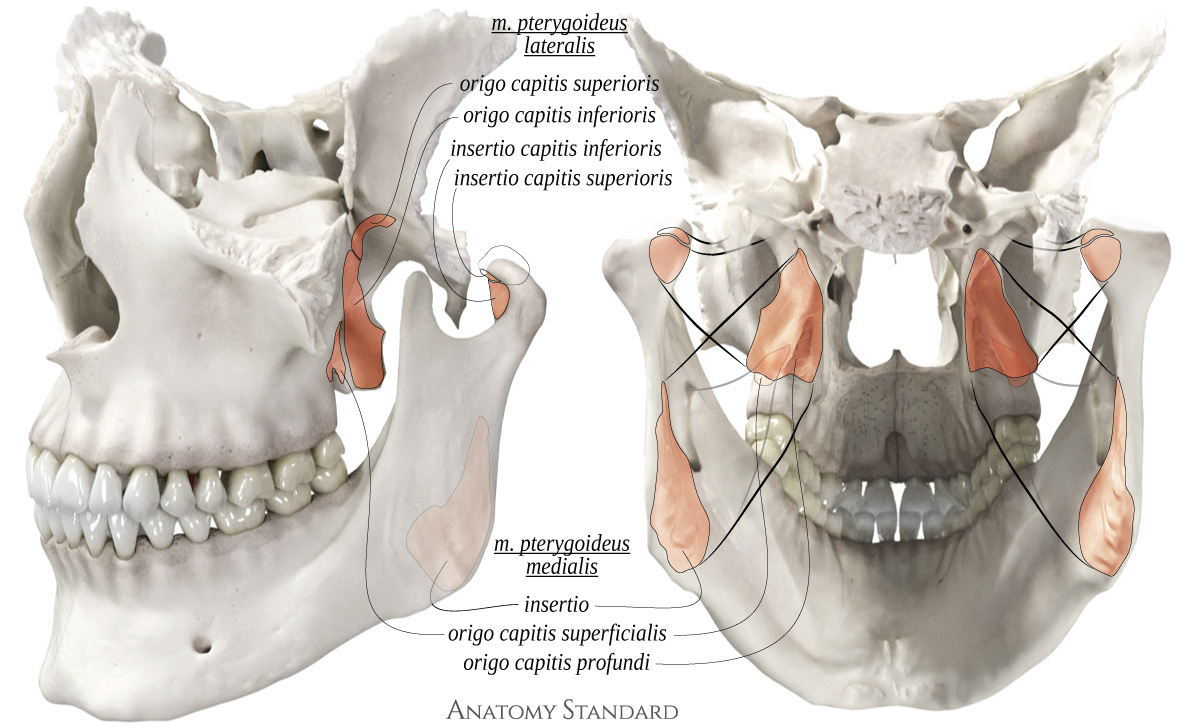
Pterygoid muscles, origo and insertio: posterior and lateral view. The medial and lateral pterygoid muscles are attached to the pterygoid process of the sphenoid bone. Latin labels.
Image (CC BY-NC) and description retrieved from Anatomy Standard, page Masticatory Muscles.
Image (CC BY-NC) and description retrieved from Anatomy Standard, page Masticatory Muscles.
Anatomical structures in item:
Uploaded by: rva
Netherlands, Leiden – Leiden University Medical Center, Leiden University
Musculus pterygoideus lateralis
Musculus pterygoideus medialis
Caput superius musculus pterygoidei lateralis
Caput inferius musculus pterygoidei lateralis
Creator(s)/credit: Jānis Šavlovskis MD, PhD, Assistant Professor; Kristaps Raits, 3D generalist
Requirements for usage
You are free to use this item if you follow the requirements of the license:  View license
View license If you use this item you should credit it as follows:
- For usage in print - copy and paste the line below:
- For digital usage (e.g. in PowerPoint, Impress, Word, Writer) - copy and paste the line below (optionally add the license icon):
"Anatomy Standard - Pterygoid muscles, origo and insertio: posterior and lateral view - Latin labels" at AnatomyTOOL.org by Jānis Šavlovskis and Kristaps Raits, license: Creative Commons Attribution-NonCommercial


Comments