
nid: 63791
Additional formats:
None available
Description:
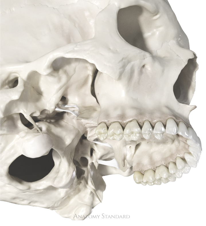
Ligamentum pterygospinale and ligamentum pterygoalare: inferolateral view. The location of these ligaments relative to the foramen ovale has significant clinical importance. The mandibular nerve passes through the foramen ovale and, branching out, comes into contact with the pterygospinal and pterygoalar ligament, passing either between the ligaments or through openings formed by the ligaments and skull. Version without labels.
Image and description retrieved from Anatomy Standard, page Cranial Syndesmoses.
Image and description retrieved from Anatomy Standard, page Cranial Syndesmoses.
Anatomical structures in item:
Uploaded by: rva
Netherlands, Leiden – Leiden University Medical Center, Leiden University
Foramen ovale
Ala major ossis sphenoidalis
Os sphenoidale
Ligamentum pterygospinale
Spina ossis sphenoidalis
Lamina lateralis processi pterygoideus ossis sphenoidalis
Creator(s)/credit: Jānis Šavlovskis MD, PhD, Assistant Professor; Kristaps Raits, 3D generalist
Requirements for usage
You are free to use this item if you follow the requirements of the license:  View license
View license If you use this item you should credit it as follows:
- For usage in print - copy and paste the line below:
- For digital usage (e.g. in PowerPoint, Impress, Word, Writer) - copy and paste the line below (optionally add the license icon):
"Anatomy Standard - Ligamentum pterygospinale and ligamentum pterygoalare: inferolateral view - no labels" at AnatomyTOOL.org by Jānis Šavlovskis and Kristaps Raits, license: Creative Commons Attribution-NonCommercial


Comments