nid: 63815
Additional formats:
None available
Description:
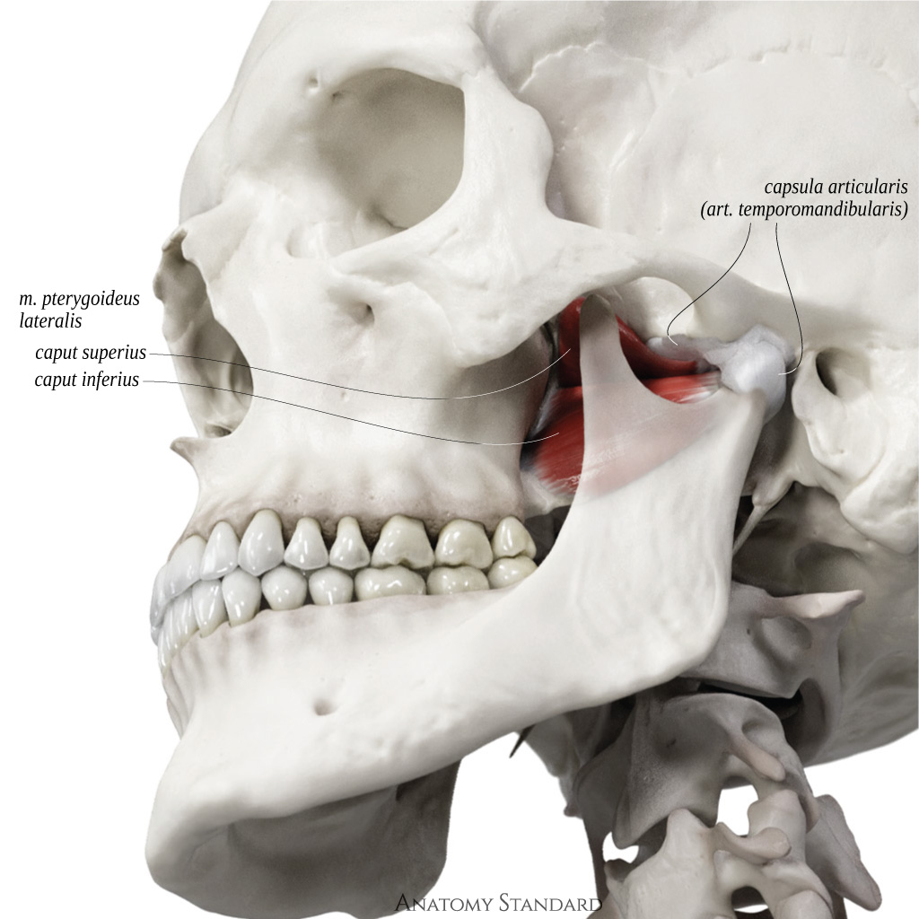
Lateral pterygoid muscle: lateral view. The medial pterygoid muscle has two heads: the upper and lower head. Upper head has its origin on the infratemporal surface of the great wing of the sphenoid bone; its insertion on the articular capsule of the TMJ and the TMJ articular disc. The function of the upper head is to pull the TMJ articular disc forward and medially. The lower head originates from the lateral surface of the lateral plate of the pterygoid process, it inserts on the fovea mandibulae. The function is to pull the condyle of mandible forward and medially. Latin labels.
Image (CC BY-NC) and description retrieved from Anatomy Standard, page Masticatory Muscles.
Image (CC BY-NC) and description retrieved from Anatomy Standard, page Masticatory Muscles.
Anatomical structures in item:
Uploaded by: rva
Netherlands, Leiden – Leiden University Medical Center, Leiden University
Musculus pterygoideus lateralis
Caput superius musculus pterygoidei lateralis
Caput inferius musculus pterygoidei lateralis
Articulatio temporomandibularis
Creator(s)/credit: Jānis Šavlovskis MD, PhD, Assistant Professor; Kristaps Raits, 3D generalist
Requirements for usage
You are free to use this item if you follow the requirements of the license:  View license
 View license
 If you use this item you should credit it as follows:
- For usage in print - copy and paste the line below:
- For digital usage (e.g. in PowerPoint, Impress, Word, Writer) - copy and paste the line below (optionally add the license icon):
"Anatomy Standard - Lateral pterygoid muscle: lateral view - Latin labels" at AnatomyTOOL.org by Jānis Šavlovskis and Kristaps Raits, license: Creative Commons Attribution-NonCommercial


Comments