
nid: 63310
Additional formats:
None available
Description:
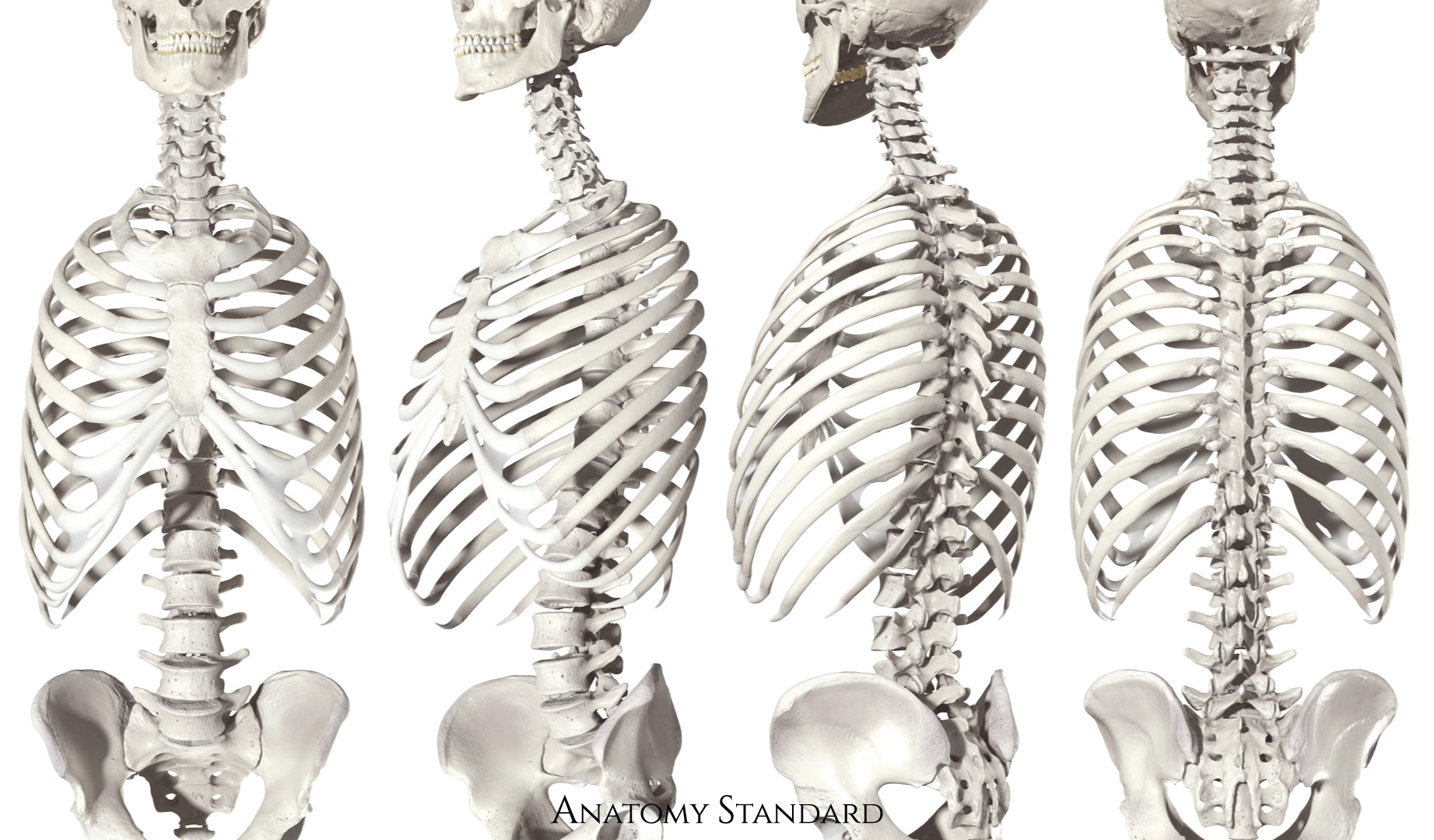
Thoracic skeleton from different views. This image shows the thoracic skeleton from anterior, lateroanterior, lateroposterior, and posterior. The thoracic skeleton consists of the twelve thoracic vertebrae, the ribs with their cartilages, and the sternum. No labels.
Image retrieved from Anatomy Standard, page Thorax.
Image retrieved from Anatomy Standard, page Thorax.
Anatomical structures in item:
Uploaded by: rva
Netherlands, Leiden – Leiden University Medical Center, Leiden University
Skeleton thoracis
Cavea thoracis
Sternum
Costae [I-XII]
Cartilago costalis
Vertebrae thoracicae (TI-TXII)
Thorax
Creator(s)/credit: Jānis Šavlovskis MD, PhD, Assistant Professor; Kristaps Raits, 3D generalist
Requirements for usage
You are free to use this item if you follow the requirements of the license:  View license
View license If you use this item you should credit it as follows:
- For usage in print - copy and paste the line below:
- For digital usage (e.g. in PowerPoint, Impress, Word, Writer) - copy and paste the line below (optionally add the license icon):
"Anatomy Standard - Drawing Thoracic skeleton from different views - no labels" at AnatomyTOOL.org by Jānis Šavlovskis and Kristaps Raits, license: Creative Commons Attribution-NonCommercial


Comments