
nid: 63883
Additional formats:
None available
Description:
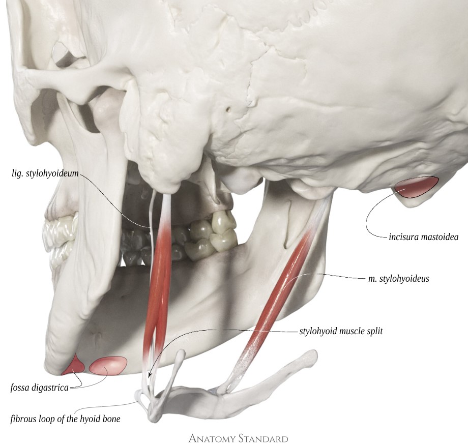
Stylohyoid muscle and insertion/origin of digastric muscle: posterolateral view. The stylohyoid muscle splits near its attachment to the hyoid bone, allowing the posterior belly of the digastric muscle to pass through. Latin labels.
Image and description (CC BY-NC) retrieved from Anatomy Standard, page Suprahyoid Muscles.
Image and description (CC BY-NC) retrieved from Anatomy Standard, page Suprahyoid Muscles.
Anatomical structures in item:
Uploaded by: rva
Netherlands, Leiden – Leiden University Medical Center, Leiden University
Ligamentum stylohyoideum
Fossa digastrica
Musculus stylohyoideus
Incisura mastoidea
Creator(s)/credit: Jānis Šavlovskis MD, PhD, Assistant Professor; Kristaps Raits, 3D generalist
Requirements for usage
You are free to use this item if you follow the requirements of the license:  View license
View license If you use this item you should credit it as follows:
- For usage in print - copy and paste the line below:
- For digital usage (e.g. in PowerPoint, Impress, Word, Writer) - copy and paste the line below (optionally add the license icon):
"Anatomy Standard Drawing - Stylohyoid muscle and insertion/origin of digastric muscle: posterolateral view - Latin labels" at AnatomyTOOL.org by Jānis Šavlovskis and Kristaps Raits, license: Creative Commons Attribution-NonCommercial


Comments