
nid: 62950
Additional formats:
None available
Description:
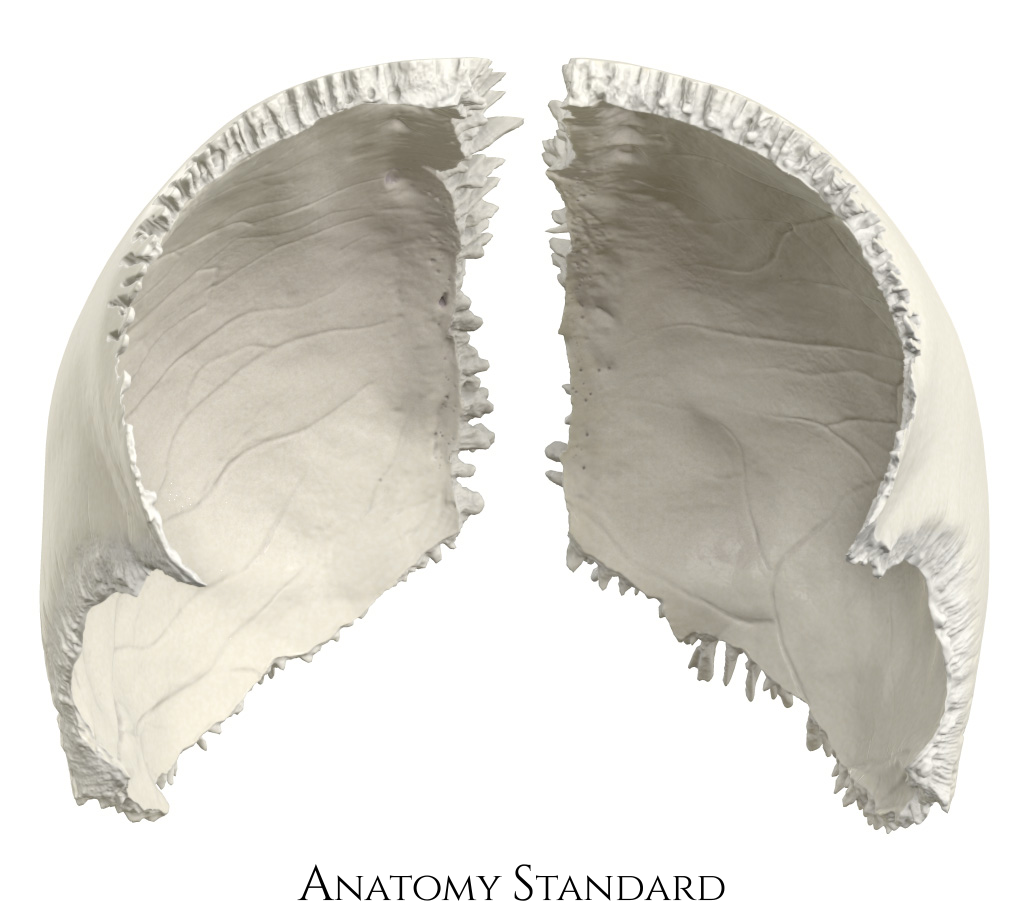
Parietal bone: anterior view. The right and left parietal bone (os parietale) are shown from anterior. In this image, they are seperated. Version without labels.
Image retrieved from Anatomy Standard.
Image retrieved from Anatomy Standard.
Anatomical structures in item:
Uploaded by: rva
Netherlands, Leiden – Leiden University Medical Center, Leiden University
Os parietale
Tuber parietale
Sulcus sinus sigmoidei (Os parietale)
Sulcus sinus sagittalis superioris
Creator(s)/credit: Jānis Šavlovskis MD, PhD, Assistant Professor; Kristaps Raits, 3D generalist
Requirements for usage
You are free to use this item if you follow the requirements of the license:  View license
View license If you use this item you should credit it as follows:
- For usage in print - copy and paste the line below:
- For digital usage (e.g. in PowerPoint, Impress, Word, Writer) - copy and paste the line below (optionally add the license icon):
"Anatomy Standard - Drawing Parietal bone: anterior view - no labels" at AnatomyTOOL.org by Jānis Šavlovskis and Kristaps Raits, license: Creative Commons Attribution-NonCommercial


Comments