
nid: 63103
Additional formats:
None available
Description:
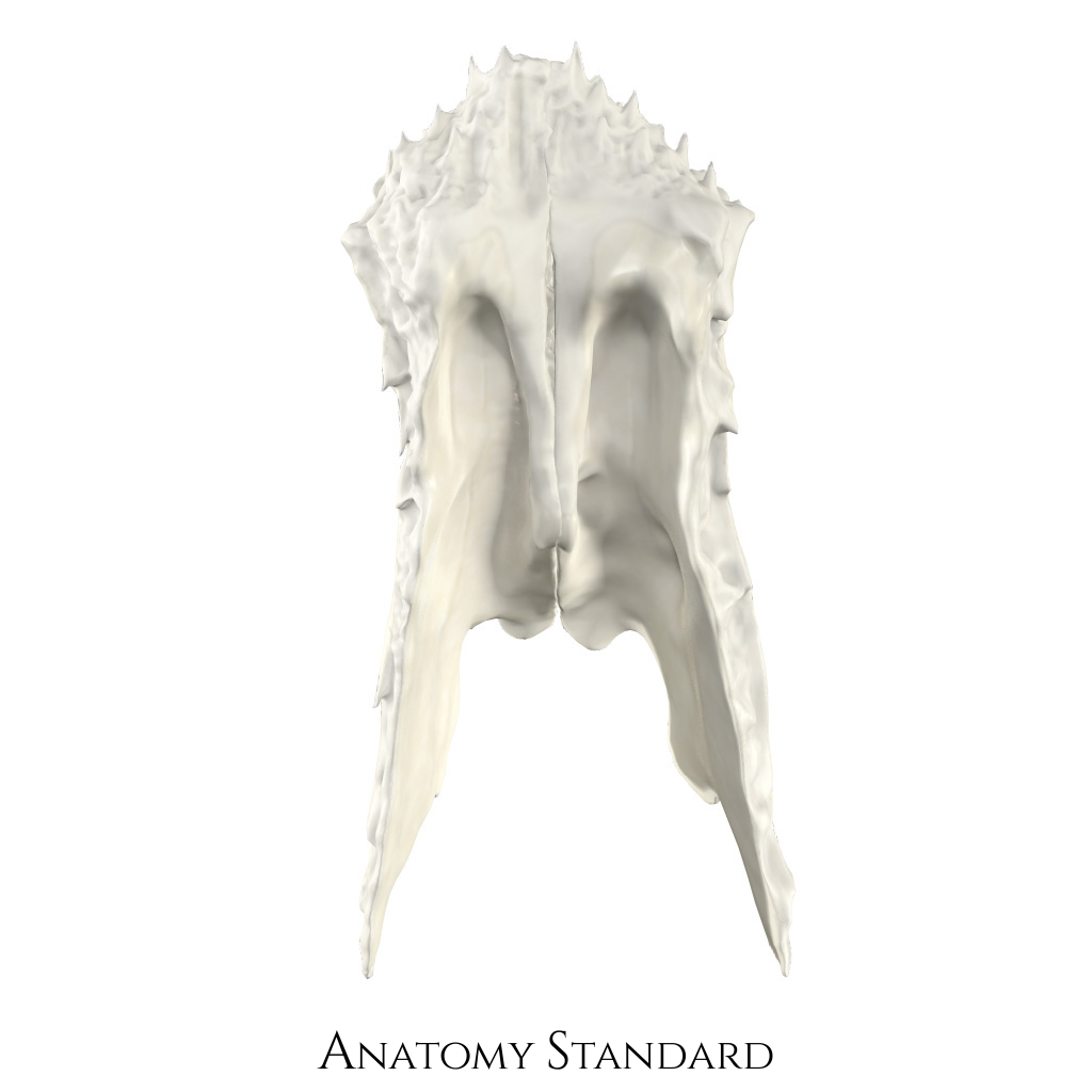
Nasal bone: posterior view. The image shows the dorsal aspect of nasal bones. Note the medial ridges that continue into the septum nasi, exactly into spina nasalis ossis frontalis and lamina perpendicularis ossis ethmoidalis. Version without labels.
Image and description retrieved from Anatomy Standard.
Image and description retrieved from Anatomy Standard.
Anatomical structures in item:
Uploaded by: rva
Netherlands, Leiden – Leiden University Medical Center, Leiden University
Os nasale
Sulcus ethmoidalis
Creator(s)/credit: Jānis Šavlovskis MD, PhD, Assistant Professor; Kristaps Raits, 3D generalist
Requirements for usage
You are free to use this item if you follow the requirements of the license:  View license
View license If you use this item you should credit it as follows:
- For usage in print - copy and paste the line below:
- For digital usage (e.g. in PowerPoint, Impress, Word, Writer) - copy and paste the line below (optionally add the license icon):
"Anatomy Standard - Drawing Nasal bone: posterior view - no labels" at AnatomyTOOL.org by Jānis Šavlovskis and Kristaps Raits, license: Creative Commons Attribution-NonCommercial


Comments