
nid: 63197
Additional formats:
None available
Description:
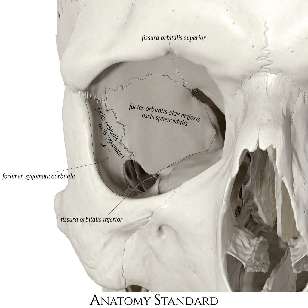
Lateral wall of the orbit. The orbit is the skull's compartment containing the eyeball, extraocular muscles, lacrimal gland, ophthalmic blood vessels, and multiple cranial nerves. This image shows the lateral wall of the orbit. Latin labels.
The anatomy of the other three walls of the orbit can be found here.
Image and description retrieved from Anatomy Standard.
The anatomy of the other three walls of the orbit can be found here.
Image and description retrieved from Anatomy Standard.
Anatomical structures in item:
Uploaded by: rva
Netherlands, Leiden – Leiden University Medical Center, Leiden University
Orbita
Paries lateralis orbitae
Fissura orbitalis superior
Facies orbitalis alaris majoris ossis sphenoidalis
Facies orbitalis ossis zygomatici
Foramen zygomaticoorbitale
Fissura orbitalis inferior
Creator(s)/credit: Jānis Šavlovskis MD, PhD, Assistant Professor; Kristaps Raits, 3D generalist
Requirements for usage
You are free to use this item if you follow the requirements of the license:  View license
View license If you use this item you should credit it as follows:
- For usage in print - copy and paste the line below:
- For digital usage (e.g. in PowerPoint, Impress, Word, Writer) - copy and paste the line below (optionally add the license icon):
"Anatomy Standard - Drawing Lateral wall of the orbit - Latin labels" at AnatomyTOOL.org by Jānis Šavlovskis and Kristaps Raits, license: Creative Commons Attribution-NonCommercial


Comments