
nid: 63156
Additional formats:
None available
Description:
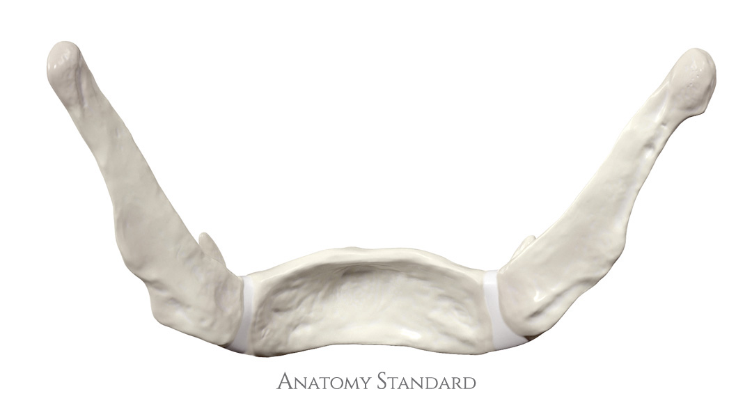
Hyoid bone: posterior view. The hyoid bone (os hyoideum) is a bone in the anterior midline of the neck and is an integral part of the larynx. No labels.
Image retrieved from Anatomy Standard.
Image retrieved from Anatomy Standard.
Anatomical structures in item:
Uploaded by: rva
Netherlands, Leiden – Leiden University Medical Center, Leiden University
Os hyoideum
Cornu majus ossis hyoidei
Cornu minus ossis hyoidei
Corpus ossis hyoidei
Creator(s)/credit: Jānis Šavlovskis MD, PhD, Assistant Professor; Kristaps Raits, 3D generalist
Requirements for usage
You are free to use this item if you follow the requirements of the license:  View license
View license If you use this item you should credit it as follows:
- For usage in print - copy and paste the line below:
- For digital usage (e.g. in PowerPoint, Impress, Word, Writer) - copy and paste the line below (optionally add the license icon):
"Anatomy Standard - Drawing Hyoid bone: posterior view - no labels" at AnatomyTOOL.org by Jānis Šavlovskis and Kristaps Raits, license: Creative Commons Attribution-NonCommercial


Comments