
nid: 63374
Additional formats:
None available
Description:
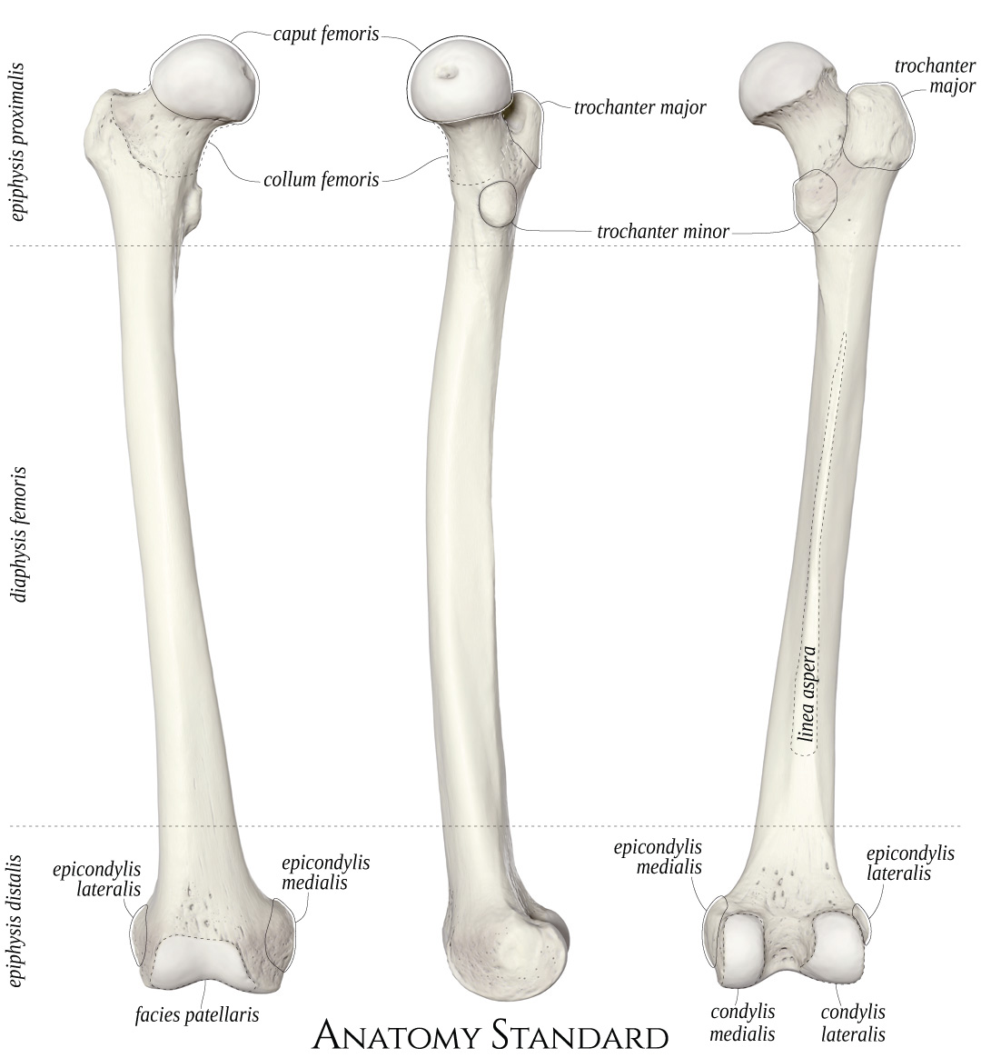
Femur: anterior, medial and posterior view. The os femoris (thigh bone) articulates proximally with the hip bone (hip joint) and distally with the tibia (knee joint). This image shows the right femur from anterior, medial and posterior. Latin labels.
Image and description retrieved from Anatomy Standard, page Femur.
Image and description retrieved from Anatomy Standard, page Femur.
Anatomical structures in item:
Uploaded by: rva
Netherlands, Leiden – Leiden University Medical Center, Leiden University
Femur
Epiphysis
Caput femoris
Collum femoris
Trochanter major
Trochanter minor
Epicondylus lateralis femoris
Linea aspera of diaphysis of femur
Epicondylus medialis femoris
Facies patellaris femoris
Condylus medialis femoris
Condylus lateralis femoris
Creator(s)/credit: Jānis Šavlovskis MD, PhD, Assistant Professor; Kristaps Raits, 3D generalist
Requirements for usage
You are free to use this item if you follow the requirements of the license:  View license
View license If you use this item you should credit it as follows:
- For usage in print - copy and paste the line below:
- For digital usage (e.g. in PowerPoint, Impress, Word, Writer) - copy and paste the line below (optionally add the license icon):
"Anatomy Standard - Drawing Femur: anterior, medial and posterior view - Latin labels" at AnatomyTOOL.org by Jānis Šavlovskis and Kristaps Raits, license: Creative Commons Attribution-NonCommercial


Comments