
nid: 63337
Additional formats:
None available
Description:
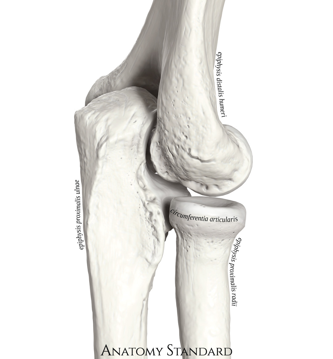
Bones of the elbow joint. An oblique postero-lateral view. The elbow joint includes three bones enclosed into one capsule. The articulation between humerus and ulna provides the flexion and extension of the forearm, whereas the axial rotation of the radius is the base of the pronation-supination motion. Latin labels.
Image and description retrieved from Anatomy Standard, page Ulna.
Image and description retrieved from Anatomy Standard, page Ulna.
Anatomical structures in item:
Uploaded by: rva
Netherlands, Leiden – Leiden University Medical Center, Leiden University
Ulna
Radius
Humerus
Articulatio cubiti
Circumferentia articularis capitis radii
Olecranon
Crista musculi supinatoris
Creator(s)/credit: Jānis Šavlovskis MD, PhD, Assistant Professor; Kristaps Raits, 3D generalist
Requirements for usage
You are free to use this item if you follow the requirements of the license:  View license
View license If you use this item you should credit it as follows:
- For usage in print - copy and paste the line below:
- For digital usage (e.g. in PowerPoint, Impress, Word, Writer) - copy and paste the line below (optionally add the license icon):
"Anatomy Standard - Drawing Bones of the elbow joint - Latin labels" at AnatomyTOOL.org by Jānis Šavlovskis and Kristaps Raits, license: Creative Commons Attribution-NonCommercial


Comments