nid: 63172
Additional formats:
None available
Description:
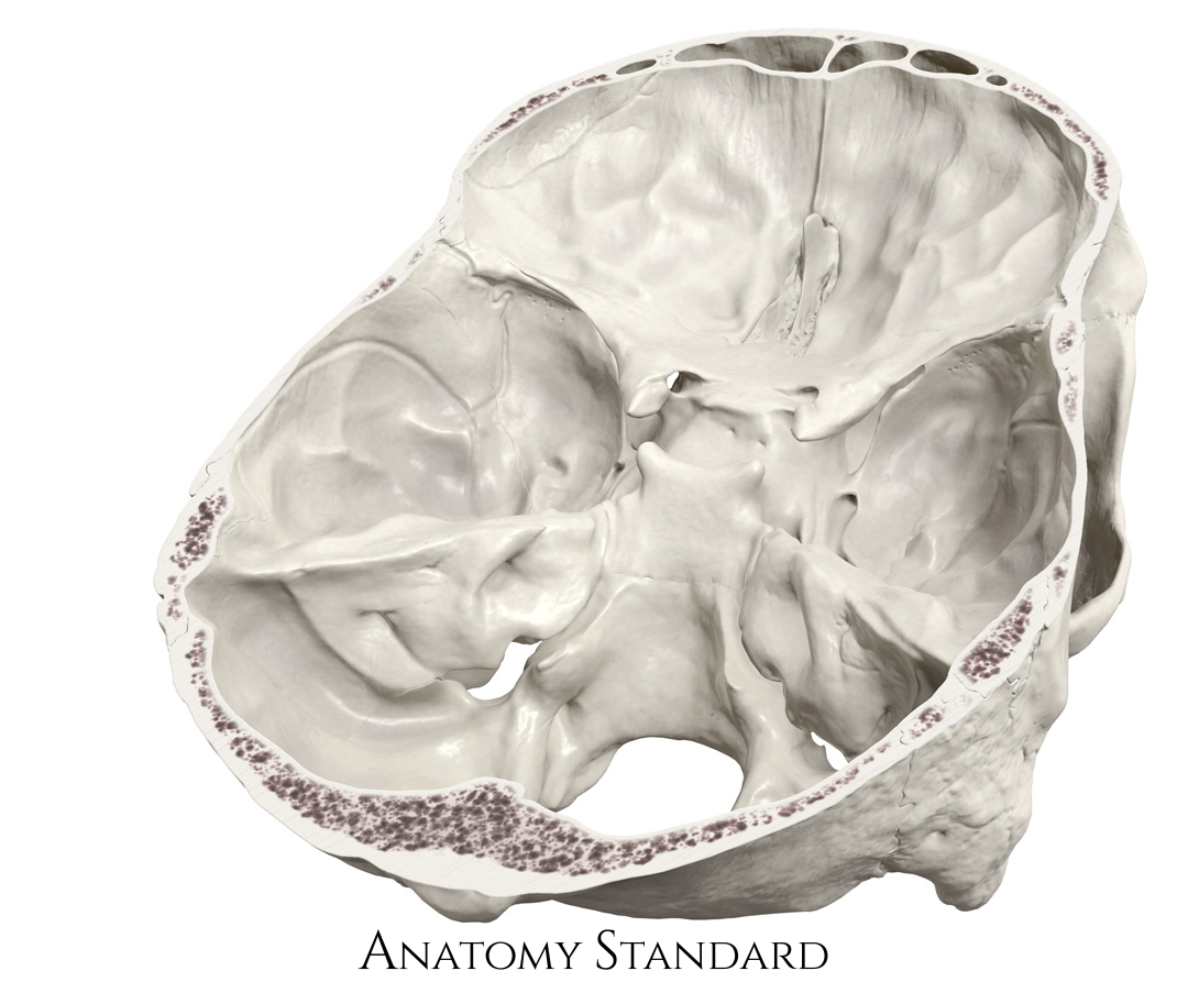
Basis cranii interna: dorsolateral view. This image demonstrates the internal surface of the base of the skull (basis cranii interna). Some important structures like the jugular foramen and the internal acoustic meatus can only be properly seen from the skull's dorsal aspect. Note also the optic canal position relative to the border between the anterior and middle cranial fossa Version without labels.
Image and description retrieved from Anatomy Standard.
Image and description retrieved from Anatomy Standard.
Anatomical structures in item:
Uploaded by: rva
Netherlands, Leiden – Leiden University Medical Center, Leiden University
Basis cranii
Basis cranii interna
Canalis opticus
Sulcus sinus petrosi inferioris
Porus acusticus internus
Meatus acusticus internus
Sulcus sinus sigmoidei
Foramen jugulare
Canalis nervi hypoglossi
Foramen rotundum
Fissura orbitalis superior
Sella turcica
Creator(s)/credit: Jānis Šavlovskis MD, PhD, Assistant Professor; Kristaps Raits, 3D generalist
Requirements for usage
You are free to use this item if you follow the requirements of the license:  View license
 View license
 If you use this item you should credit it as follows:
- For usage in print - copy and paste the line below:
- For digital usage (e.g. in PowerPoint, Impress, Word, Writer) - copy and paste the line below (optionally add the license icon):
"Anatomy Standard - Drawing Basis cranii interna: dorsolateral view - no labels" at AnatomyTOOL.org by Jānis Šavlovskis and Kristaps Raits, license: Creative Commons Attribution-NonCommercial


Comments