
nid: 59467
Additional formats:
None available
Description:
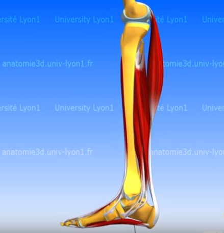
In this video the organization and function of the foot muscles is shown and explained. The intrinsic and extrinsic muscles of the foot can be distinguished, these muscles are short and located in the foot respectively long and with their origin on the leg.
This video is in the "3D Anatomy Lyon" series from the Université Claude Bernard in Lyon, France. NOTE: THIS VIDEO IS UNDER A NON-DERIVATIVE LICENSE. THIS MEANS THAT IF YOU REMIX, TRANSFORM, OR BUILD UPON THE MATERIAL, YOU MAY NOT DISTRIBUTE THE MODIFIED MATERIAL.
This video is in the "3D Anatomy Lyon" series from the Université Claude Bernard in Lyon, France. NOTE: THIS VIDEO IS UNDER A NON-DERIVATIVE LICENSE. THIS MEANS THAT IF YOU REMIX, TRANSFORM, OR BUILD UPON THE MATERIAL, YOU MAY NOT DISTRIBUTE THE MODIFIED MATERIAL.
Anatomical structures in item:
Uploaded by: rva
Netherlands, Leiden – Leiden University Medical Center, Leiden University
Pes
Musculus tibialis anterior
Musculus extensor hallucis longus
Musculus extensor digitorum longus
Musculus tibialis posterior
Musculus flexor hallucis longus
Musculus flexor digitorum longus
Musculus triceps surae
Musculus gastrocnemius
Musculus soleus
Plantaris
Musculus fibularis longus
Musculus fibularis brevis
Musculus adductor hallucis
Musculus abductor metatarsi quinti
Creator(s)/credit: Olivier Rastello, multimedia expert; Patrice Thiriet PhD, anatomist, project leader of Anatomie 3D Lyon; Nora van Reeth, project manager; Christophe Batier, technical director; Cédric Carré
Requirements for usage
You are free to use this item if you follow the requirements of the license:  View license
View license If you use this item you should credit it as follows:
- For usage in print - copy and paste the line below:
- For digital usage (e.g. in PowerPoint, Impress, Word, Writer) - copy and paste the line below (optionally add the license icon):
"3D Anatomy Lyon: Organization and fuction of the foot muscles - video of 3D model" at AnatomyTOOL.org by Olivier Rastello, Patrice Thiriet, Nora van Reeth et al, license: Creative Commons Attribution-NonCommercial-NoDerivs. video in the "Anatomie 3D Lyon" series at https://www.youtube.com/channel/UC9LucUID-BUjL_c8oAT3vHQ/
"3D Anatomy Lyon: Organization and fuction of the foot muscles - video of 3D model" by Olivier Rastello, Patrice Thiriet, Nora van Reeth et al, license: CC BY-NC-ND. video in the "Anatomie 3D Lyon" series at https://www.youtube.com/channel/UC9LucUID-BUjL_c8oAT3vHQ/


Comments