
nid: 59410
Additional formats:
None available
Description:
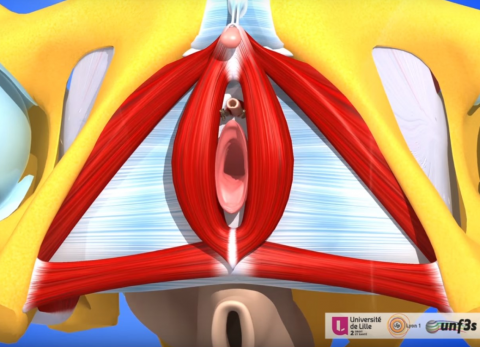
In this video the muscles and fascias of the female perineum are built up step by step with a 3D model. The division of the perineum in urogenital perineum and anal perineum is shown. Also characteristics of the external female genitals are shown, such as the paraurethral and major vestibular glands, the clitoris and the hymen. Note some deviations from the common anatomical interpretations. 1) The posterior boundary of the perineum is incorrectly stated to be the sacroiliac ligament. This should be the sacrotuberous ligament. 2) The urogenital triangle and anal triangle are called urogenital perineum and anal perineum in the video. 3) The video appears to follow closer the clinical interpretation of perineum -only the superficial structures- than the common anatomical interpretation - the whole compartment inferior of the pelvic diaphragm. This video is in the "3D Anatomy Lyon" series from the Université Claude Bernard in Lyon, France. NOTE: THIS VIDEO IS UNDER A NON-DERIVATIVE LICENSE. THIS MEANS THAT IF YOU REMIX, TRANSFORM, OR BUILD UPON THE MATERIAL, YOU MAY NOT DISTRIBUTE THE MODIFIED MATERIAL.
Anatomical structures in item:
Uploaded by: opgobee
Netherlands, Leiden – Leiden University Medical Center, Leiden University
Pelvis
Musculus levator ani
Diaphragma pelvis
Vulva
Clitoris
Perineum
Crus clitoridis
Bulbus vestibuli
Glans clitoridis
Glandula vestibularis major
Ductus paraurethrales
Musculus transversus perinei superficialis
Musculus transversus perinei profundus
Musculus bulbospongiosus
Musculus ischiocavernosus
Musculi perinei
Fascia perinei
Corpus perineale
Labium majus pudendi
Labium minus pudendi
Hymen
Corpus clitoridis
Ligamentum suspensorium clitoridis
Musculus sphincter ani externus
Regio urogenitalis
Regio analis
Creator(s)/credit: Géraldine Giraudet MD, gynaecologist-obstetrician; Chrystèle D. Rubod MD, gynaecologist-obstetrician; Prof. Michel Cosson MD, gynaecologist-obstetrician; Patrice Thiriet PhD, anatomist, project leader of Anatomie 3D Lyon; Olivier Rastello, multimedia expert
Requirements for usage
You are free to use this item if you follow the requirements of the license:  View license
View license If you use this item you should credit it as follows:
- For usage in print - copy and paste the line below:
- For digital usage (e.g. in PowerPoint, Impress, Word, Writer) - copy and paste the line below (optionally add the license icon):
"3D Anatomy Lyon: The female perineum - video of 3D model" at AnatomyTOOL.org by Géraldine Giraudet, Chrystèle D. Rubod, Michel Cosson et al, license: Creative Commons Attribution-NonCommercial-NoDerivs. video in the "Anatomie 3D Lyon" series at https://www.youtube.com/channel/UC9LucUID-BUjL_c8oAT3vHQ/
"3D Anatomy Lyon: The female perineum - video of 3D model" by Géraldine Giraudet, Chrystèle D. Rubod, Michel Cosson et al, license: CC BY-NC-ND. video in the "Anatomie 3D Lyon" series at https://www.youtube.com/channel/UC9LucUID-BUjL_c8oAT3vHQ/


Comments