
nid: 59462
Additional formats:
None available
Description:
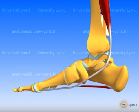
In this video the muscles of the posterior compartment of the leg are shown and explained. A good basis to start studying the muscles of the leg. The fibularis brevis muscle is accidentally labeled as popliteal muscle at 0:17, at 0:32 the popliteal is labeled correctly. This video is in the "3D Anatomy Lyon" series from the Université Claude Bernard in Lyon, France. NOTE: THIS VIDEO IS UNDER A NON-DERIVATIVE LICENSE. THIS MEANS THAT IF YOU REMIX, TRANSFORM, OR BUILD UPON THE MATERIAL, YOU MAY NOT DISTRIBUTE THE MODIFIED MATERIAL.
Anatomical structures in item:
Uploaded by: rva
Netherlands, Leiden – Leiden University Medical Center, Leiden University
Crus
Musculus popliteus
Musculus flexor hallucis longus
Musculus tibialis posterior
Musculus flexor digitorum longus
Fossa poplitea
Musculus popliteus
Ligamentum cruciatum posterius
Calcaneus
Hallux
Musculus fibularis brevis
Creator(s)/credit: Olivier Rastello, multimedia expert; Patrice Thiriet PhD, anatomist, project leader of Anatomie 3D Lyon; Nora van Reeth, project manager; Christophe Batier, technical director
Requirements for usage
You are free to use this item if you follow the requirements of the license:  View license
View license If you use this item you should credit it as follows:
- For usage in print - copy and paste the line below:
- For digital usage (e.g. in PowerPoint, Impress, Word, Writer) - copy and paste the line below (optionally add the license icon):
"3D Anatomy Lyon: Deep posterior compartment of the leg - video of 3D model" at AnatomyTOOL.org by Olivier Rastello, Patrice Thiriet, Nora van Reeth et al, license: Creative Commons Attribution-NonCommercial-NoDerivs. video in the "Anatomie 3D Lyon" series at https://www.youtube.com/channel/UC9LucUID-BUjL_c8oAT3vHQ/
"3D Anatomy Lyon: Deep posterior compartment of the leg - video of 3D model" by Olivier Rastello, Patrice Thiriet, Nora van Reeth et al, license: CC BY-NC-ND. video in the "Anatomie 3D Lyon" series at https://www.youtube.com/channel/UC9LucUID-BUjL_c8oAT3vHQ/


Comments